Medical expert of the article
New publications
Exophthalmos
Last reviewed: 04.07.2025

All iLive content is medically reviewed or fact checked to ensure as much factual accuracy as possible.
We have strict sourcing guidelines and only link to reputable media sites, academic research institutions and, whenever possible, medically peer reviewed studies. Note that the numbers in parentheses ([1], [2], etc.) are clickable links to these studies.
If you feel that any of our content is inaccurate, out-of-date, or otherwise questionable, please select it and press Ctrl + Enter.

Causes of exophthalmos
The direction of proptosis may indicate the underlying disease. For example, lesions located within the muscular infundibulum, such as cavernous hemangiomas or optic nerve tumors, result in axial proptosis, while lesions located outside the muscular infundibulum typically result in displaced proptosis, the direction of which is determined by the location of the lesion.
Symptoms of exophthalmos
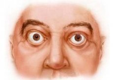
Exophthalmos is classified as axial, unilateral or bilateral, symmetrical or asymmetrical, and is often permanent. Severe exophthalmos may interfere with eyelid closure, leading to exposure keratopathy and corneal ulceration.
False exophthalmos (pseudoexophthalmos) can occur with facial asymmetry, unilateral enlargement of the eyeball (with high myopia or buphthalmos), unilateral retraction of the eyelid or eophthalmos on the opposite side.
Diagnosis of exophthalmos
The severity of exophthalmos is measured with a plastic ruler applied to the outer edge of the orbit or with a Heriel exophthalmometer equipped with mirrors in which the corneal apexes are visible and a special scale is applied. Ideally, measurements should be taken in two positions: looking up and looking down. Values greater than 20 mm indicate the presence of exophthalmos, and a difference in eye protrusion of 2 mm is suspicious regardless of the absolute value of exophthalmos. Exophthalmos is divided into mild (21-23 mm), moderate (24-27 mm) and severe (28 mm and more). The width of the palpebral fissure and any lagophthalmos should be taken into account.
What do need to examine?
How to examine?
Who to contact?
Treatment of exophthalmos
The approach to treating exophthalmos is controversial. Some suggest early decompression surgery, while others advise resorting to surgery only after conservative methods of treating exophthalmos have proven ineffective or insufficient.
- Systemic use of steroids is indicated for rapidly increasing exophthalmos with pain syndrome in the edema stage, if there are no contraindications (for example, tuberculosis or peptic ulcer).
- Oral prednisolone (initial dose 60-80 mg daily). Discomfort, chemosis, and periorbital edema usually subside within 48 hours, then the steroid dose is gradually reduced. Maximum results are seen after 2-8 weeks. Ideally, steroid therapy should be completed within 3 months, although low-dose maintenance therapy may be necessary for a long time;
- intravenous methylrednisolone (0.5 g in 200 ml isotonic saline over 30 min). Repeat after 48 h. This can be effective and is usually recommended for compression optic neuropathy. However, there is a risk of cardiovascular complications, so therapeutic monitoring is necessary.
- Radiotherapy is an alternative when steroids are contraindicated or ineffective. The effect usually appears within 6 weeks and is maximal by 4 months.
- Combination treatment with radiotherapy, azathioprine, and low-dose prednisolone may be more effective than using steroids and radiotherapy alone.
- Surgical decompression can be used as a primary method or when conservative methods are ineffective (for example, with disfiguring exophthalmos in the fibrosis stage). Decompression, which is often performed endoscopically, is of the following types:
- two-wall - antral-ethmoidal decompression with removal of sections of the lower and posterior part of the inner wall. This achieves a reduction in exophthalmos by 3-6 mm;
- three-wall - antral-ethmoidal decompression with removal of the outer wall. The effect is 6-10 mm;
- four-wall - three-wall decompression with removal of the outer half of the orbital vault and most of the main bone at the orbital apex. This allows to reduce exophthalmos by 10-16 mm, therefore it is used in cases of severe exophthalmos.


 [
[