Medical expert of the article

New publications
Oral erythroplakia
Last reviewed: 04.07.2025

All iLive content is medically reviewed or fact checked to ensure as much factual accuracy as possible.
We have strict sourcing guidelines and only link to reputable media sites, academic research institutions and, whenever possible, medically peer reviewed studies. Note that the numbers in parentheses ([1], [2], etc.) are clickable links to these studies.
If you feel that any of our content is inaccurate, out-of-date, or otherwise questionable, please select it and press Ctrl + Enter.
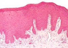
Erythroplakia of the oral cavity is a persistent red spot. It has no clinical manifestations. In most cases, the neoplasm is characterized as epithelial dysplasia. The whole danger is that the tumor can become malignant. Usually, everything is localized on the mandibular transitional fold in the oropharynx, tongue and vestibule of the mouth. The patient does not complain about anything, nothing bothers him. It appears in people over 55 years old, both women and men.
ICD-10 code
Thanks to a specially created international classification, each disease was assigned its own unique code. Thus, diseases of the oral cavity, salivary glands and jaw are designated as K00-K14. K00 Disorders of development and eruption of teeth. Only retained and impacted teeth are excluded.
- K01 Retained and impacted teeth. Only those that are malpositioned in relation to adjacent teeth are excluded.
- K02 Dental caries.
- K03 Other diseases of hard tissues of teeth. Excludes bruxism, dental caries and teeth grinding NEC.
- K04 Diseases of the pulp and periapical tissues. K05 Gingivitis and periodontal diseases.
- K06 Other changes of gingiva and edentulous alveolar ridge. Atrophy of the edentulous alveolar ridge and gingivitis are excluded.
- K07 Dentofacial anomalies [including malocclusions]. Excludes atrophy and hypertrophy of hemiface (Q67.4) unilateral condylar hyperplasia or hypoplasia (K10.8).
- K08 Other changes in teeth and their supporting apparatus.
- K09 Cysts of oral region, not elsewhere classified. Also includes lesion with histologic features of aneurysmal cyst and other fibro-osseous lesion. Excludes radicular cyst (K04.8).
- K10 Other diseases of the jaws.
- K11 Disease of the salivary glands.
- K12 Stomatitis and related lesions. Excludes decomposing ulcer of the mouth (A69.0) cheilitis (K13.0) gangrenous stomatitis (A69.0) gingivostomatitis due to herpes virus.
- K13 Other diseases of the lips and oral mucosa. This includes changes in the epithelium of the tongue. Completely excludes: certain changes in the gingiva and edentulous alveolar ridge (K05-K06) cysts of the oral region (K09) diseases of the tongue (K14) stomatitis and related lesions (K12).
- K14 Diseases of tongue. Excludes erythroplakia, focal epithelial, hyperplasia of tongue (K13.2) leukedema, leukoplakia, hairy leukoplakia (K13.3) macroglossia (congenital) (Q38.2) submucous fibrosis of tongue (K13.5)
Causes of oral erythroplakia
People who have bad habits are at risk. This concerns alcohol and tobacco. Moreover, smoking is not necessary, it is enough to just use chewing tobacco, the result will be similar. The combination of alcohol and tobacco increases the risk of developing a cancerous tumor several times. Most often, men suffer from this pathology, but due to the sharp increase in women smokers, there is no particular difference between the sexes. Everyone is susceptible to tumor formation.
Cigarettes, pipes and cigars significantly increase the risk of malignant neoplasms in the human oral cavity. This occurs especially often at the point where the cigarette touches the lips. At first, a spot appears that looks like a mole. A biopsy can determine whether the spot is malignant or not.
Broken teeth can also provoke the development of erythroplakia. Dentures and fillings have a similar effect. There is a risk of developing a malignant tumor. People who have previously suffered from this pathology, the risk of recurrence is high.
 [
[ Pathogenesis
Among all oral lesions, cancer is the most common. The formation can be both benign and malignant. The beginning of the process can be explained. Thus, the consideration is conducted from the point of view of the polyetiological theory of origin. Any damage, be it mechanical irritation, temperature, chemical or biological factors - all this negatively affects the oral cavity. Unsatisfactory oral condition, poor hygiene and other "little things" can aggravate the situation. All this, individually or in combination, leads to problems with the oral cavity. In almost 50% of cases, erythroplakia occurs against the background of carious teeth. Orthopedic structures cause tumors in 10%.
As for the etiological factors, smoking, placing NASA under the tongue, chewing betel and burns with ethyl alcohol have a special influence. All this leads to damage to the oral mucosa and, as a consequence, to tumors. Constant heating and toxicity of tobacco can cause a neoplasm.
Symptoms of oral erythroplakia
The whole danger is that the person does not complain of any discomfort. Usually, erythroplakia does not manifest itself in any way until it becomes a malignant tumor. Changes can be noticed only visually. Thus, bright red spots can appear on the mucous membrane of the oral cavity. Many people do not pay attention to this, and in vain. Those who are frightened by this "symptom" go to the hospital. The doctor collects anamnesis from the patient regarding his lifestyle and the presence of bad habits.
This pathology is characterized by the presence of limited bright red spots. In addition to them, there may be small soft nodules, which cause pain when palpated. Upon careful examination of the oral cavity, several red spots can be seen in different areas. This should alert a person. This color is caused by atrophy of the mucous membrane, as a result of which the vessels located in the submucosa become visible.
Spots can be seen in the molar area and on the mucous membrane. They are usually located on the transitional fold of the lower jaw, tongue and bottom. Sometimes patients are bothered by the symptoms of both erythroplakia and leukoplakia. This condition increases the risk of developing cancer several times.
First signs
The first thing that appears is swelling and thickening of the affected area on the oral mucosa. Seals and bumps can be felt. Some areas become rough, covered with crusts and look like small erosions. Velvety white or red spots appear. They mainly appear on the inside of the oral cavity.
There is no pain or other discomfort. Unexplained bleeding in the oral cavity may begin. Numbness, loss of sensitivity of a separate area, pain - appear at later stages. More precisely, only in the case of a malignant course.
Chronic ulcers on the face, neck and mouth may indicate a problem. They may bleed slightly and not heal for 2 weeks or more. A sensation of a foreign body may appear in the throat. When chewing, discomfort, pain and a change in taste appear. All this is typical for later stages.
Consequences
The problem cannot be ignored. After all, erythroplakia is a precancerous condition. If treatment is not started in time, the tumor can become malignant. As a consequence, a fatal outcome. Treatment must be correct and include several methods. Usually, the pathology is eliminated with the help of radiation and chemotherapy. Everything is eliminated surgically.
The consequences are affected not only by the treatment provided, but also by the recovery period. It is important to correctly diagnose the pathology, determine the size of the tumor, its location and damage to adjacent tissues.
After surgery, many patients experience weakness and general malaise. This is quite normal. It goes away on its own in about a few weeks. Timely treatment guarantees success. If you ignore erythroplakia, it becomes malignant and the only consequence can be a fatal outcome.
Complications
Without proper treatment, complications can be irreparable. It is worth noting that the problem is eliminated by chemotherapy, radiation therapy and surgery. It is impossible to eliminate the pathology with drugs. Moreover, without high-quality treatment, erythroplasty of the oral cavity will take a malignant form.
Correct elimination consists of making a diagnosis. Thus, it is important to determine not only the location of the spot, but also its size, as well as the factors that led to such an event. Then treatment is prescribed. Usually it is complex and begins with the removal of that very formation. It will not be possible to leave it or remove the spot with medications.
If a person starts treatment on time, there will be no complications. There can be only two. The first option is the transition to a malignant form, the second option is a fatal outcome. The importance and promptness of actions will help a person avoid both pronounced symptoms and serious consequences.
Diagnostics
The attending physician should carefully examine the oral cavity. Particular attention is paid to the sublingual area. Additionally, the examination requires a visit to a specialized medical institution. The examination is performed using a special mirror, as well as a lamp. To ensure that there are no ulcers in the throat, you will have to use an endoscope with a thin flexible tube and a light bulb at the end.
In order to make a diagnosis, a biopsy will have to be performed. To do this, the doctor removes a tiny area and examines it under a microscope. This procedure is performed exclusively under general anesthesia. Therefore, the person will have to spend some time in the hospital. After this, further examination is carried out.
To assess a person's condition, it is necessary to take a blood test and also to conduct an X-ray of the chest organs. It is important to examine the oral cavity for metastases. Based on the results of the analysis, high-quality treatment is prescribed. In some cases, the lesion affects the bones, as well as individual parts of the facial part of the skull. To determine the presence of a problem, it is necessary to conduct an orthopantogram.
Magnetic resonance imaging is also widely used. It allows for detailed examination of tissues and organs. Before the procedure, the patient is asked to remove all metal objects and jewelry from their body.
Computer tomography plays a special role. A series of X-ray images will allow you to study the oral cavity layer by layer and get acquainted with all the pathologies in it. Before the procedure, a person should not drink or eat for 4 hours. In case of complications, a bone scan is performed. This will allow you to see pathological changes in the facial parts of the skull.
Tests
In order to study the affected area, a biopsy will have to be performed. The procedure involves taking tissue from the tumor site and examining it in detail under a microscope. During the procedure, the person is under anesthesia. The obtained material must be sent for histological examination. Experienced laboratory doctors examine the tissue area and make their conclusions. Usually, if a tumor is present, changes characteristic of a certain type of neoplasm are detected.
In addition to a biopsy, a person must take a blood test. Thanks to it, it becomes possible to study the affected area and identify changes in it at the cellular level. Here, pathological changes are also seen, concerning enzymes, metabolites and some tumor markers. These tests are taken together. Thanks to them, it is possible to get a complete picture of what is happening and prescribe high-quality treatment.
Instrumental diagnostics
This diagnostic method includes several main directions. So, first of all, a person needs to undergo nasopharyngoscopy. Thanks to this procedure, it becomes possible to more thoroughly examine the back wall of the oral cavity for pathological changes.
Pharyngoscopy and laryngoscopy are widely used. These procedures are necessary for examining the mucous membrane of the larynx and trachea. In case of possible complications with damage to the facial part of the skull, X-ray of the bones is performed. It allows identifying the main foci of tumor growth.
Scintigraphy. The procedure is a study using radioactive isotopes. This is an informative examination that helps to identify the presence of metastases in bone tissue.
Computer, magnetic resonance and positron emission tomography. These diagnostic methods help to clarify the nature of the neoplasm, as well as the degree of damage. All of the above procedures can be used both separately and in combination.
Differential diagnostics
This method of research includes several methods. Thus, in addition to using instrumental diagnostics, it is worth taking a sample of the affected tissue and donating blood for analysis. Thus, a person who has suspicions of erythroplakia of the oral cavity must undergo a biopsy procedure. It is an excision of a small affected area. For this, a person must be subjected to general anesthesia. The excised area is submitted for histological examination. It is examined under a microscope for the presence of pathological processes.
In addition to the biopsy, you will also have to take a blood test. Any changes in the body are immediately visible in the blood. Thus, the material can be used to study the cellular composition and make sure that there is a change in the indicators. Usually, the neoplasm changes enzymes, metabolites and some tumor markers. Thanks to these two procedures, and in combination with instrumental diagnostics, you can not only make a correct diagnosis and prescribe high-quality treatment.
Who to contact?
Treatment of oral erythroplakia
Surgical, radiation and medicinal methods are used to eliminate the problem. Much depends on the person's condition and the affected area. The doctor chooses the treatment method based on the diagnostic data obtained.
Surgical treatment. Various methods are used to remove the tumor. Usually, the movable part of the oral cavity and oropharynx is removed. In this case, the bones are not affected. In case of damage to the facial part of the jaw, the affected area is sawed out. More details about the surgical method of treatment will be described below.
Radiation therapy. This method is the main treatment for people with tumors in the oral cavity and oropharynx. The procedure is used together with surgery, the main purpose of which is to eliminate the affected area. External irradiation is often used. Treatment should be carried out 5 times a week for 5-7 weeks. Some patients are prescribed brazitherapy. It is internal irradiation. Specialists insert special metal rods containing radioactive material into the tumor and areas located near it. When the person is discharged home, the rods are removed. In most cases, both external and internal irradiation are actively used. The method has side effects. They include reddening of the skin, dryness, pain in the throat, as well as weakness and loss of taste. Complications can include damage to the thyroid gland and blood vessels.
Chemotherapy. This method involves the use of special antitumor drugs. This method can be used together with surgical removal and radiation therapy. This will not only eliminate the tumor itself, but also avoid complications. The drugs used are Cisplatin, Fluorouracil, Docetaxel, Paclitaxel and Gemcitabine. Detailed information about them will be provided below. Chemotherapy can cause a number of side effects. These include nausea, vomiting, general weakness and loss of appetite. The person is plagued by rapid fatigue, and there is also an increased susceptibility to infection.
Drug treatment
No medications are used independently. Most of them are part of a comprehensive treatment of the problem, namely one method - chemotherapy. Cisplatin, Fluorouracil, Docetaxel, Paclitaxel and Gemcitabine are widely used.
- Cisplatin. The drug can be used both separately and in combination therapy. The dosage is prescribed individually and depends on the patient's condition. Usually 20 mg per square meter is administered. The administration is carried out daily for 5 days, then 3 weeks are left between courses. The drug has a number of side effects, it can disrupt the liver and kidneys, lead to nausea, vomiting and general malaise. Contraindications: hypersensitivity, ulcer, pregnancy, liver and kidney dysfunction.
- Fluorouracil. It is used to eliminate tumors, malignant type. The dosage is prescribed by the attending physician. 15 mg per kilogram of weight is enough. The introduction is carried out over 4 hours. The frequency of use and duration are selected individually. Contraindications: hypersensitivity, severe diarrhea, pregnancy, infectious diseases, liver and kidney dysfunction. Side effects: nausea, vomiting, liver and kidney dysfunction, confusion.
- Docetaxel. The drug is used exclusively intravenously. 0.74 mg per ml is sufficient. The administration is carried out over 4 hours. The duration of use of the drug is discussed individually. Contraindications: hypersensitivity, liver failure, breastfeeding and pregnancy. Side effects: nausea, vomiting, fatigue, liver and kidney dysfunction, erythema, skin itching.
- Paclitaxel. The dosage is prescribed individually depending on the person's condition, as well as the tumor. Contraindications: hypersensitivity, pregnancy, lactation period and neutropenia. Side effects: anemia, nausea, vomiting, diarrhea, allergic reactions, necrosis.
- Gemcitabine. The drug is administered intravenously and drip for 30 minutes. It is advisable to use it no more than once a week. The course duration is 3 weeks. Repeated administration no earlier than after 7 days. Contraindications: hypersensitivity, pregnancy and lactation. Side effects: headache, nausea, weakness, vomiting, diarrhea, constipation, stomatitis.
Folk remedies
In case of cancerous and precancerous formations, the use of traditional medicine is a bit inappropriate. The problem needs to be dealt with more professionally. But despite this, there are several basic methods of eliminating a tumor using traditional medicine.
- Recipe 1. Take 10 grams of dry crushed chamomile flowers, marshmallow root and juniper berries. For a better effect, add a head of garlic. Mix all the ingredients together, chop the garlic. Then pour a liter of cold water over everything and bring to a boil. Then simmer for an hour over low heat. Cool the resulting solution and rinse your mouth with it.
- Recipe 2. You need to take 100 grams of garlic juice and a couple of tablespoons of walnut leaves ground into powder. For maximum effect, nettle is also used. The resulting ingredients are mixed and 500 ml of liquid honey is added to them. The resulting mixture allows you to restore the body's strength.
- Recipe 3. Take garlic and squeeze the juice out of it. The first five days take 10 drops, the next 5 days take 20 drops. Thus, the dosage is brought to 2 tablespoons per day.
Herbal treatment
Herbs are folk medicine. Today, it is used quite often. But in the presence of serious inflammatory processes and cancerous tumors, it is inappropriate to resort to its help. More precisely, it can be used only in combination with other methods.
Recipe 1. Take 100 grams of calendula petals and pour half a liter of alcohol (60 degrees) over them. The resulting tincture is sent to a dark place for 10 days. The contents should be shaken periodically. After the specified time has passed, strain the tincture and take one teaspoon per day. In addition, you should eat 200 grams of carrot gruel. Season it with 3-5 cloves of garlic, you can also add onions.
Recipe 2. You need to take calendula flowers and bedstraw grass. To prepare the remedy, take 2 tablespoons of the mixture and pour 500 ml of water. Cook everything over low heat for 5 minutes. Then cool and strain. Take the remedy ¼ cup up to 4 times a day 15 minutes before meals.
Homeopathy
Homeopathic remedies are also widely used, although they are not always of particular importance. It is still recommended to resort to the help of traditional medicine. Despite this, the most basic homeopathic remedies will be presented below.
- Carcinosin. It is used exclusively in a dilution of 200 or 1000. It can be used once a week or a month. Other drugs are used in parallel.
- Conium. The remedy has already demonstrated its effectiveness. It is used exclusively in dilutions of 200 or 1000.
- Arsenicum. Effective for burning sensation. Potassium cyanatum 30, 200 - useful for cancer of the tongue. It is especially often used for neuralgia of the facial nerves.
- Hydrastis. This tincture is effective for uterine erythroplakia. The solution can be used for douching. It is actively used for oral cavity lesions. The remedy helps relieve pain. It can be used no more than 2 times a week.
- Carbo animalis 30 - when pus breaks through. Aconite radix is used to relieve pain, 1 or 2 drops. This method is used until the pain syndrome completely disappears.
- Phosphorus. Widely used for tumors in the oral cavity, on the lips and cheeks. The patient experiences a strong feeling of thirst and requires ice water.
There are many other medications that are used depending on the symptoms that appear. An effective remedy can only be selected with a homeopathic doctor.
Surgical treatment
Various surgeries can be used to perform this technique. In this case, the location of the tumor, the stage of development, and the need for restorative interventions are taken into account.
In patients with a tumor in the oral cavity, removal is performed without capturing bone tissue. If mobility is significantly limited, the affected area is removed along with part of the jaw. Jaw damage can be seen on an X-ray.
If the tumor is on the lip, a special surgical micrographic method is used. In this case, the tumor is removed layer by layer using a microscope. This will allow the tumor to be completely removed while preserving normal lip tissue.
Malignant tumors are usually "famous" for affecting the lymph nodes located in the neck. Therefore, the removal procedure involves the removal of suspicious lymph nodes. The scope of the operation depends entirely on the spread of the tumor. Sometimes it is necessary to remove muscles, nerves and blood vessels.
This method can lead to complications. Thus, numbness of the ear, drooping of the lower lip and difficulty raising the arms above the head are possible. This is due to nerve damage. Sometimes breathing difficulties occur.
Prevention
Many cases of oral tumor development can be prevented. To do this, you just need to eliminate known negative factors. Thus, tobacco and smoking pose a particular risk. In most cases, they lead to the development of tumors. After all, the lips, oral cavity and mucous membranes are constantly exposed to the negative effects of nicotine. The best solution is to get rid of the bad habit.
It is important to understand that smoking and drinking alcohol increase the risk of developing erythroplakia several times. Therefore, it is worth reviewing your own life. The risk of the problem is high. It is important to avoid exposure to the sun at its peak. The negative impact of ultraviolet radiation can lead to lip cancer.
It is enough to simply eliminate bad habits and start eating special products. Thus, vegetables, fruits and products from coarse grains can reduce the risk of developing pathology several times.
Forecast
After the tumor removal procedures are completed, a person may experience some problems with speech and swallowing. It is possible to eliminate all of this, but not on your own. You should seek help from a nutritionist and speech therapist. They will conduct an examination, listen to the patient, and prescribe appropriate procedures based on the data obtained.
People who have had a malignant neoplasm are at risk of recurrence of the problem. Relapse can occur within 2 years from the day of tumor removal. Therefore, patients should always be under close medical supervision.
Patients who have undergone radiation therapy risk reducing the level of hormones produced by the thyroid gland. To fully get rid of the problem, it is worth seeking help from an endocrinologist and undergoing a course of therapy prescribed by him.
It has been proven that patients who have had cancer have a risk of recurrence of the tumor. A particular risk is observed when drinking alcohol and smoking. Therefore, it is better to get rid of these bad habits.