Medical expert of the article

New publications
Epiphyseolysis in children
Last reviewed: 29.06.2025

All iLive content is medically reviewed or fact checked to ensure as much factual accuracy as possible.
We have strict sourcing guidelines and only link to reputable media sites, academic research institutions and, whenever possible, medically peer reviewed studies. Note that the numbers in parentheses ([1], [2], etc.) are clickable links to these studies.
If you feel that any of our content is inaccurate, out-of-date, or otherwise questionable, please select it and press Ctrl + Enter.
Displacement or detachment of the neocostal epiphyseal plate (sprout cartilage) - epiphyseolysis in children - can be detected in cases of tubular bone fractures in the metaepiphyseal region where this cartilaginous plate is located.
This is only seen in childhood and adolescence when bony growth continues, while in adults the epiphyseal plates undergo ossification, that is, they are replaced by mature bone, leaving an epiphyseal scar. [1]
Epidemiology
According to clinical statistics, epiphyseolysis occurs in almost 15% of tubular bone fractures in childhood. Epiphyseal plate fractures are twice as common in boys as in girls, because bone growth ends earlier in girls (accelerated skeletal maturation is due to estrogen).
The most frequent localization of epiphysiolysis is noted in fractures of the lower radius of the forearm and the distal tibia of the tibia.
Causes of the epiphyseolysis in children
Causes of epiphyseolysis - injuries to bones and joints in children, which can occur as a result of traffic accidents, hitting a limb, falling while running, jumping, cycling (skateboarding, skating); due to excessive and frequently repeated loads on bones during sports training.
Fractures of the tubular bones of the skeleton in children and adolescents involving the metaepiphyseal zones and growth plates (physis), which are located between the expanded part of the bone body (metaphysis) and the end of the bone (epiphysis) and provide longitudinal growth of the limbs, are called Salter-Harris fractures. There are five types of such fractures.
A Type I fracture is a transverse fracture through the growth plate, affecting the cartilage but not affecting the bone. The injury may cause separation of the epiphysis or rounded end of the bone from the bone shaft. Type II fracture - fracture through an area most of the growth plate and metaphysis, the horizontal fracture line ascends upward at an angle, affecting the areas above the growth plate; separation of the metaphyseal fragment may occur.
A type III fracture crosses the epiphyseal plate toward the epiphysis (with preservation of the metaphysis) and may involve the joint, while type IV fractures pass vertically through the growth zone, metaphysis, and epiphysis. The rarest type V fracture is a compression fracture of the epiphyseal plate.
Also read the publication - fractures
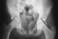
Slipped epiphysis of the femoral head with abnormal angle of the epiphysis relative to the metaphysis - juvenile epiphyseolysis of the femoral head - may not be associated with acute trauma, but develop as an osteochondropathy or orthopedic deformity as a result of compression and local shear forces in children with severe secondary hyperparathyroidism, hypocalcemia, chronic renal failure, and severe fibrous osteitis of the adjacent metaphysis - due to changes in the structure of the growth cartilage and its partial fibrosis.
Risk factors
Orthopedic surgeons and trauma surgeons consider the risk factors for epiphyseolysis to include an increased risk of fracture in children with pathologic changes in bone structure and low bone mass.
And such a condition, defined as secondary osteoporosis, can develop due to the presence in children: hyperthyroidism, primary hyperparathyroidism, juvenile rheumatoid arthritis, hypercorticism (Cushing's syndrome), hypopituitarism (with a deficiency of somatotropin - growth hormone), diabetes mellitus, gluten enteropathy (celiac disease), hypocalcemia and vitamin D deficiency (rickets), congenital osteogenesis imperfecta, homocystinuria or bone mineral metabolism disorders in chronic kidney disease.
Pathogenesis
Considering the peculiarities of bone development and growth, the pathogenesis of epiphyseolysis in children is explained by the fact that the weakest and most vulnerable to injury areas of the immature pediatric skeleton are the epiphyseal cartilages, as they cannot fully resist shear stress in case of fractures or excessive loads.
The epiphyseal plates of long bones are translucent cartilaginous strips separating the epiphysis from the metaphysis, which are composed of chondrocytes in a collagen matrix; they undergo several stages of maturation and are replaced by osteoblasts, osteoclasts, and lamellar bone during endochondral ossification. This process is regulated not only by chondrocytes (which divide and grow by producing extracellular matrix), but also by a variety of humoral factors: growth hormone, parathormone, estrogen, cytokines, fibroblast growth factor (FGF), insulin-like growth factor (IGF-1), signaling peptides, and others.
When it enters the fracture area, a gap or cleavage forms in the sprouting cartilage, which causes damage to its structure and can impair chondrocyte function.
Symptoms of the epiphyseolysis in children
The first signs of bone fracture with capture of the growth plate are manifested by constant pain in the injured limb.
Other common symptoms include: swelling at the end of the bone, localized hyperthermia and pain when pressure is applied near the joint; hematoma; forced position of the limb; deformity of the limb; limitation of mobility - inability to bend/extend the limb.
Localization of epiphyseolysis in lower extremity bone fractures includes:
- Epiphyseolysis of the femoral head in children as a result of an intra-articular fracture of the femur, affecting its head, which is located at the upper end of the bone. Although the wavy shape of the distal femur and the presence of the mastoid bodies provide additional stability of the growth plate, there is a higher likelihood of post-traumatic bone growth arrest when it is fractured. [2]
- Epiphyseolysis of the tibia (thick tibia) in children is very often the result of trauma to the distal part of the tibia (when a plantar flexion force is applied to the supinated foot) with type II (Salter-Harris) displacement of the growth cartilage. For more information see. - epiphyseolysis of the tibia
- Epiphyseolysis of the fibula in children may occur in epiphyseal fractures of the thin lateral bone of the tibia in its lower part.
- Epiphysiolysis of the ankle joint in a child may be observed in a spiral fracture of the fibula of the lower third of the tibia (so-called Maisonneuve's fracture) with rupture of the distal interosseous syndesmosis and interosseous membrane.
- Epiphyseolysis of the ankle in children is noted with concomitant fracture of the inner ankle or rupture of the deep deltoid ligament of the ankle joint - with displacement and inclination of the talus.
- Epiphyseolysis of the heel bone in children is the result of its fracture, which most often occurs when falling from a height.
Fractures of the bones of the upper extremities are possible:
- Epiphyseolysis of the head of the humerus in children - with intra-articular fracture of the ball-shaped thickening of its upper epiphysis, fracture of the distal epiphysis and condyle head of the lower epiphysis of the humerus; [3]
- Epiphyseolysis of the cephalic eminence of the humerus in children or the small head of the humerus in cases of fracture of its distal end near the epiphysis and articulation with the ulna;
- Epiphyseolysis of the ulna in children - in metaepiphyseal fractures in the upper or lower parts of the bone.
- Epiphyseolysis of the radius in a child - with a fracture of its distal metaepiphysis or fracture of the head of the radius, which is often a consequence of a fall on the straightened arm. Fractures of both forearm bones should also be considered, especially in the
Stages of epiphyseolysis are determined by specialists depending on the angle of displacement of the sprouting cartilage: if it does not exceed 30°, the stage is considered mild; if it reaches 50°, epiphyseolysis of the middle stage is diagnosed, and the severe stage is a shift of 50° or more.
Complications and consequences
Most growth plate fractures with a mild stage of displacement heal without complications, but severe damage to the growth cartilage in young children (in the active phase of bone growth) can produce effects and complications such as:
- Shortening of the leg when its longitudinal growth stops due to premature ossification of the growth plate;
- Curvature of the limb due to the formation of a bone bridge across the fracture line with displacement. The deformity is more pronounced with severe displacement or destruction of the neocostal epiphyseal plate and can lead to functional instability of the joint and degenerative arthritis.
Poorly healing trauma to the growth plate may be complicated by avascular osteonecrosis.
Diagnostics of the epiphyseolysis in children
Visualization is the basis for the diagnosis of growth plate lesions. That's why it's used
Instrumental diagnostics: radiography of the bone in the straight and lateral projections, X-ray of the joints (arthrography).
However, unossified epiphyseal plates are not visualized by X-rays, so ultrasound, CT or MRI scans are used.
For example, a CT scan allows you to clearly see the fracture, assess the degree of joint misalignment, and plan for fixation. [4]
Differential diagnosis
The differential diagnosis should exclude osteonecrosis, osteochondroma, achondroplasia, dissecting osteochondritis, osteoblastoclastoma, fibrous osteodysplasia, bone cysts, and osteosarcoma.
Who to contact?
Treatment of the epiphyseolysis in children
The choice of treatment tactics for epiphyseolysis depends on the localization of the growth plate fracture, the stage of its displacement and the degree of deformity, the presence of bone displacement, as well as the age of the child.
Most type I and II fractures require closed repositioning and immobilization with a plaster cast. Healing of these fractures occurs within two to three weeks of injury and problems are rare, especially in areas such as the distal radius.
Type III and IV fractures involve the articular surface, so open repositioning with either external fixation - percutaneous osteosynthesis, or internal fixation is required.
Surgical treatment is performed when bone fragments are displaced and the fracture is unstable. The most common surgery is called open repositioning with internal fixation. First, the bone fragments are moved to their normal position and then the fracture is fixed (with screws, spokes, pins or plates). After surgery, a bandage is applied to protect and immobilize the injured area while it heals.
Prevention
Preventing epiphyseolysis in children is fracture prevention, which, in addition to following safety precautions, may include preventing osteoporosis in children.
Forecast
With proper treatment, most growth plate fractures heal without adverse effects, but if treatment is done improperly or not at all - complications can lead to disability in children.
Использованная литература