Medical expert of the article
New publications
Epicanthus: causes, symptoms, diagnosis, treatment
Last reviewed: 07.07.2025

All iLive content is medically reviewed or fact checked to ensure as much factual accuracy as possible.
We have strict sourcing guidelines and only link to reputable media sites, academic research institutions and, whenever possible, medically peer reviewed studies. Note that the numbers in parentheses ([1], [2], etc.) are clickable links to these studies.
If you feel that any of our content is inaccurate, out-of-date, or otherwise questionable, please select it and press Ctrl + Enter.
Epicanthus is a crescent-shaped vertical fold of skin between the upper and lower eyelids that partially covers the inner corner of the eye slit and changes its configuration, which creates a false impression of convergent strabismus.
Epicanthus is found in most children under 6 months of age, and in adults it is a characteristic feature of representatives of the Mongoloid race. Normally, epicanthus is observed in children with a flat bridge of the nose, as it develops, most of the folds gradually decrease with the child's growth and rarely remain by the age of 7. Bilateral epicanthus is a frequently observed sign of various chromosomal disorders (Down syndrome). The fold can cover the inner corner of the eye, which creates a false impression of convergent strabismus.
Symptoms of epicanthus
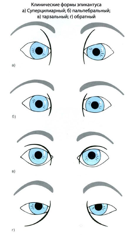
- Epicanthus palpebralis. Skin folds are symmetrically distributed between the upper and lower eyelids. Most common in Caucasians:
- Epicanthus tarsalis. Skin folds that start from the middle of the upper eyelid and extend medially to the canthus. Most common in Easterners;
- Epicanthus inversus. Skin folds begin in the lower eyelid and extend upward to the medial canthus. Often associated with blepharophimosis syndrome;
- Epicanthus superciliaris. Folds of skin originate from the eyebrow and extend downward and laterally toward the nose.

Clinical forms of epicanthus. a) Superciliary, b) palpebral, c) tarsal, d) reverse
What do need to examine?
How to examine?


 [
[