Medical expert of the article

New publications
Glandular polyp of the endometrium
Last reviewed: 08.07.2025

All iLive content is medically reviewed or fact checked to ensure as much factual accuracy as possible.
We have strict sourcing guidelines and only link to reputable media sites, academic research institutions and, whenever possible, medically peer reviewed studies. Note that the numbers in parentheses ([1], [2], etc.) are clickable links to these studies.
If you feel that any of our content is inaccurate, out-of-date, or otherwise questionable, please select it and press Ctrl + Enter.
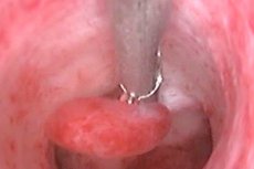
A nodular neoplasm of glandular cells with the inclusion of atypical elements is a glandular polyp. Very often, such growths appear on the mucous membrane of the uterine cavity. The growth can be spherical, branched or mushroom-shaped. It consists of a network of branching glands on the endometrium. In addition to the uterus, such neoplasias are found on the walls of the stomach and intestines.
Glandular polyps are acquired pathologies, the risk of which increases in the presence of the following factors:
- Endocrine diseases.
- Hormonal disorders.
- Hereditary predisposition.
- Inflammatory processes and diseases of the pelvic organs.
- Hormonal dysfunction during menopause.
- Long-term trauma to the mucous membrane during long-term use of an intrauterine device.
- Surgical interventions: abortion, curettage, probing of the uterine cavity.
- Immunodeficiency.
- Unstable emotional background, frequent stress.
Local intrauterine tissue proliferation can be caused by hypovitaminosis E and C, a weakened immune system, excess body weight, and intestinal diseases (colitis, Crohn's disease).
According to statistics, about 15% of cases of intrauterine neoplasms do not cause symptoms. But in most cases, women note the following signs:
- Heavy and painful menstruation.
- Delayed menstruation followed by heavy bleeding.
- Intermenstrual bleeding.
- Pulling pain in the lower abdomen.
- Purulent discharge from the genitals.
- Secondary anemia.
- Infertility.
If the glandular neoplasm is large, cramping pains appear. Large growths are the cause of infertility and have a high risk of malignancy. If the size is more than 2 cm, the risk of its degeneration is 10%. In this case, formations on a broad base often become malignant.
To diagnose the disease, ultrasound examination, hysteroscopy and histology of the complete scraping of the mucous membrane of the uterine cavity are performed. Treatment is surgical. The operation is performed under local or general anesthesia. The patient is prescribed a course of drug therapy to restore hormonal levels.
Particular attention should be paid to preventive measures. For early diagnosis of any changes, it is necessary to undergo regular gynecological examinations, promptly treat inflammatory and any other diseases. It is also necessary to take care of strengthening the immune system.
Glandular fibrous polyp of the endometrium
A small, limited growth of the mucous membrane of the uterine walls, consisting of connective tissue elements and glandular structures, is a glandular-fibrous polyp of the endometrium. Its development occurs in the direction of the uterine cavity. The growth structure is divided into a body and a stalk. Most often, it is localized at the bottom of the uterus, and when it reaches large sizes, it blocks the cervical canal. In this case, the neoplasm is benign.
The main causes of glandular fibrous neoplasia of the uterus:
- Dysfunction of the ovaries. Failure of the production of sex hormones leads to a decrease in progesterone production and an increase in estrogen synthesis. Because of this, a focus of inflammation is formed in the endometrium, which is not rejected during menstruation, but on the contrary increases in size.
- Dysfunction of the adrenal glands.
- Long-term use of an intrauterine device.
- Miscarriages and abortions.
- Diseases that cause metabolic disorders in the body. The risk of polyposis is increased in women with hypertension, diabetes, and obesity.
In most cases, the pathological condition is asymptomatic, which leads to late diagnosis and treatment. But there are a number of signs that allow you to suspect polyps in the uterus:
- Menstrual cycle disorders.
- Bloody discharge not associated with menstruation.
- Heavy menstruation.
- Pain in the lower abdomen after intercourse.
- Increased volume of normal vaginal discharge.
During the diagnostics, the gynecologist asks the patient about painful symptoms, conducts a visual examination on the chair, and an ultrasound examination of the uterus. Treatment is surgical. The neoplasia is removed by surgical resection with scraping of the mucous membrane of the uterine cavity. The operation is performed under the control of hysteroscopy.
To reduce the risk of relapse and possible complications, the tissue removal site is treated with liquid nitrogen. Hormonal therapy is administered to restore the menstrual cycle and prevent relapses.
Glandular cystic polyp of the endometrium
Another type of endometrial neoplasm is glandular cystic polyps. Such a growth contains glands of different shapes and lengths, the stroma at the base is denser, fibrous. The glands are unevenly located with cystically stretched lumens. According to histology, proliferative glandular epithelium alternates with non-functioning.
Pathological proliferation of the glandular layer with the simultaneous formation of cysts occurs due to the following factors:
- Hormonal disorders.
- Endocrine disorders and diseases.
- Inflammatory and infectious processes of the internal genital organs.
- Gynecological diseases: polycystic disease, endometriosis, uterine fibroids.
- Dysfunction of the adrenal and thyroid glands.
- Hypertension, obesity.
- Genetic predisposition.
A glandular cystic local intrauterine formation may proceed unnoticed. But as its tissues grow, the following symptoms appear:
- Bloody discharge from the genital tract before or after menstruation.
- Menstrual cycle disorders.
- Minor pain in the lower abdomen.
- Dizziness and general weakness.
- Discomfort during sexual intercourse.
- Long-term unsuccessful attempts to get pregnant.
The presence of one or more of the above symptoms is a reason to seek immediate medical help. The sooner the diagnosis and treatment are carried out, the lower the risk of complications.
Conservative therapy is powerless, so surgical intervention is indicated for treatment. In case of glandular cystic hyperplasia, hysteroscopy is performed. The growth is completely removed and a thorough scraping of the organ mucosal surface is performed. To reduce the risk of relapse, the site of the removed tissue is treated with liquid nitrogen. The tissues obtained as a result of the operation are sent for histology. If atypical cells are detected in the analysis, then a gynecologist-oncologist is engaged in further treatment of the patient.
Basal type glandular polyp of the endometrium
According to histology, the uterus consists of several structures:
- The inner layer is the endometrium.
- The middle layer is the myometrium.
- External - serous membrane or perimetrium.
Each of these structures has substructures. For example, the endometrium consists of a functional and basal layer (adjacent to the myometrium). The thickness of the basal layer is 1-1.5 mm, it consists of connective tissue elements, contains parts of the myometrium glands, blood vessels and nerve endings. At the same time, it is insensitive to hormonal changes and is not rejected during menstruation. The proliferation of its cells restores the normal structure of the endometrium.
But under the influence of certain factors, significant changes occur. For example, with hyperestrogenism, the cells of the basal layer regenerate very quickly, which leads to its thickening, i.e. hyperplasia. Against this background, glandular polyps of the endometrium of the basal layer very often occur. They can be asymptomatic, but as they grow, they cause menstrual irregularities and other painful symptoms.
Treatment of such local intrauterine formations is surgical. The patient undergoes hysteroresectoscopy with histological examination of tissues. If the growth contains atypical cells, then further treatment is carried out by a gynecologist-oncologist.
 [ 9 ], [ 10 ], [ 11 ], [ 12 ], [ 13 ], [ 14 ], [ 15 ], [ 16 ]