Medical expert of the article

New publications
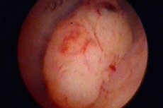
Atypical and malignant endometrial polyps
Last reviewed: 08.07.2025

All iLive content is medically reviewed or fact checked to ensure as much factual accuracy as possible.
We have strict sourcing guidelines and only link to reputable media sites, academic research institutions and, whenever possible, medically peer reviewed studies. Note that the numbers in parentheses ([1], [2], etc.) are clickable links to these studies.
If you feel that any of our content is inaccurate, out-of-date, or otherwise questionable, please select it and press Ctrl + Enter.
Under the influence of certain factors, any neoplasm that has arisen in the body can take a malignant form. This also applies to intrauterine polypous growths. They are most often diagnosed in older women (menopause, postmenopause).
Diagnosis of malignant endometrial polyps is performed using histological examination. The tissues collected during hysteroscopy are sent for analysis. The following precancerous conditions can be identified based on the results of histology:
- Adenomatous neoplasm.
- Glandular polyp with cell proliferation.
- Carcinoma in situ (early forms of oncology).
According to the conducted research, the main reason for malignancy of benign growths is genetic and hormonal disorders. In the first case, it is a hereditary predisposition. The risk of developing cancer increases under the influence of the following factors:
- Endocrine pathologies.
- Gynecological diseases.
- Inflammatory processes in the endometrium.
- Tumor lesions of the uterus and its appendages.
Uterine polyps are especially dangerous during menopause and those that arise against the background of endocrine disorders. The first signs of intrauterine pathologies include menstrual cycle disorders. This manifests itself in irregular or heavy periods. There may be nagging pain in the lower abdomen, increased vaginal discharge, weakness and general fatigue.
The pathology is diagnosed using transvaginal ultrasound. The diagnosis is confirmed by hysteroscopy followed by histological examination of the collected tissues.
The treatment is performed by a gynecologist-oncologist. The malignant neoplasm is removed and the uterine cavity is scraped. The patient is then prescribed drug therapy to correct the hormonal background. Particular attention is paid to preventive measures: normalization of body weight, refusal of abortions, timely treatment of gynecological and any other diseases of the body, regular examinations by a gynecologist.
Atypical endometrial polyp
A neoplasm of the endometrium with abnormal structures, which arose as a result of tumor transformations and inflammatory processes of tissues, is an atypical polyp. The presence of atypical cells indicates the risk of malignant transformation of the growth.
An atypical (adenomatous) polyp can form from any type of tissue. Transformation is associated with the action of certain factors. Depending on the degree of structural change in the mucous membrane, two types of atypical hyperplasia are distinguished:
- Simple - in histological analysis, an increased number of glandular and stromal elements, but without structurally altered endometrium. The glands have increased mitotic activity. These may be glandular or glandular-cystic polyps.
- Complex – endometrium with pronounced proliferation of the glandular component. There are signs of atypia at the tissue and cellular level. Invasion of the basement membrane of glandular structures is absent. Histology indicates an accumulation of abnormal cells, loss of polarity of the glands. Cellular atypia is characterized by proliferation and distortion of the shape of the glands with infiltrates and endometrial stroma.
Atypical changes are a precancerous condition, i.e. an intermediate position between the usual forms of glandular hyperplasia and oncology. The malignant potential of atypical polyps is 30-50%.
 [
[ Endometrial polyp without atypia
According to histological classification, hyperplastic processes of the endometrium are divided into the following groups:
- Glandular cystic hyperplasia.
- Endometrial polyps: glandular, cystic, fibrous.
- Atypical hyperplasia (polyps, adenomatosis, etc.).
The first two groups are the background for endometrial cancer and occur in 2-4% of cases. Atypical processes are characterized by a violation of cellular differentiation within the epithelial layer, i.e. they are precancer.
An endometrial polyp without atypia indicates a benign proliferation of mucosal tissue. Histological examination of the neoplasm does not reveal malignant cells. The safest growths include anomalies of the functional layer of the mucosa of a glandular or fibrous nature.
For the treatment of polyps without atypia, surgical removal and restoration of hormonal balance with the help of medications are indicated.