Medical expert of the article

New publications
Placental polyp of the endometrium
Last reviewed: 04.07.2025

All iLive content is medically reviewed or fact checked to ensure as much factual accuracy as possible.
We have strict sourcing guidelines and only link to reputable media sites, academic research institutions and, whenever possible, medically peer reviewed studies. Note that the numbers in parentheses ([1], [2], etc.) are clickable links to these studies.
If you feel that any of our content is inaccurate, out-of-date, or otherwise questionable, please select it and press Ctrl + Enter.
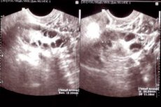
A specific intrauterine formation formed from the remains of placental tissue after a miscarriage, abortion or childbirth is a placental endometrial polyp. Blood clots settle on the remaining tissues of the placenta, which is tightly attached to the uterus, and form a benign growth. According to medical statistics, about 10% of women face this problem.
The main cause of placental growth is the retention of chorionic tissue in the uterine cavity. There are also a number of factors that increase the risk of developing this pathology:
- Death of the fetus in the womb.
- Medical or classic abortion, miscarriage with incomplete removal of the site of attachment of the fertilized egg.
- Incomplete removal of the placenta after cesarean section.
- Incorrect management of the postpartum period.
In some cases, a placental neoplasm occurs during pregnancy, but is not dangerous for the mother and fetus. It is formed from the tissue of the placenta or fetal membranes and is removed from the body during childbirth.
The symptoms of the disease are rather vague, since many women believe that bloody discharge after an abortion or childbirth is normal. Therefore, the main sign of the pathology is spotting or bloody discharge over a long period of time. In addition, the patient complains of pain in the lower abdomen, deterioration of general health, pale skin, increased body temperature, itching and burning in the genital area.
If the above symptoms appear, you should seek medical help. This is because without treatment, pathological signs progress and cause life-threatening complications:
- Inflammation of the uterine mucosa.
- Ovarian dysfunction.
- Development of infection, sepsis.
- Female infertility.
- Anemia.
For diagnosis, the doctor collects anamnesis, determines whether there was a pregnancy, miscarriage or abortion, curettage of the uterine cavity. During a gynecological examination, the uterus is enlarged and painful, an elastic seal is palpated. An ultrasound is performed to confirm the growth. Treatment consists of removing the neoplasm. For this, curettage or vacuum aspiration is used. Removal with a laser is also possible.
Decidual polyp of the endometrium
This type of neoplasm occurs during pregnancy. After conception, the woman's hormonal background undergoes changes, due to which the endometrium begins to actively grow, that is, its decidualization occurs. During this period, a small growth (decidual polyp) may form, protruding into the lumen of the cervical canal or beyond.
Causes of pathology:
- Hormonal disorders.
- Cervical trauma.
- Weakened immune system.
- Increased estrogen levels.
- Endocrine pathologies.
- Urogenital infections.
According to medical statistics, about 22% of pregnant women experience polypoid lesions of the uterine mucosa. In this case, women are diagnosed with two types of neoplasms:
- Decidual pseudopolyps.
- True polyps with decidualization.
The neoplasm contains a large number of blood vessels, so at the slightest trauma (sexual intercourse, medical manipulations) it begins to bleed. This in turn is dangerous due to the development of infectious and inflammatory processes.
Symptoms of decidual intrauterine formation:
- Pain and spasms in the lower abdomen.
- Change in color and intensity of discharge.
- Elevated body temperature.
- Bloody discharge.
For diagnostics, a colposcopic examination of the cervix and cervical canal is performed. This procedure is painless and does not take much time. The woman is also prescribed a set of general clinical and bacteriological laboratory tests.
The detected polyps are differentiated from papilloma of the cervix, injured tissues and prolapsing glands. To determine the condition of the growth, a smear is taken for oncocytology. After the final diagnosis is made, a treatment plan is drawn up.
As a rule, decidual polyp of the endometrium does not require therapy and goes away on its own after the hormonal background is restored. At the same time, such growths do not interfere with the birth process. Therefore, the gynecologist monitors the neoplasm and the state of the internal flora of the vagina. If the growth bleeds, there are ulcers or destructive changes on its surface, it provokes spasms and increases the tone of the uterus, then it is removed.
 [ 1 ], [ 2 ], [ 3 ], [ 4 ], [ 5 ]
Chorionic endometrial polyp
Placental (chorionic) polyp is a part of the placental tissues tightly attached to the endometrium in the uterus. The neoplasm can occur after childbirth or abortion. Blood clots are layered on the placental tissues, forming a seal. As it grows, it is covered by the placental membrane.
The main causes of chorionic neoplasm:
- Incomplete removal of the placenta after cesarean section.
- Frozen pregnancy.
- Late-term abortion.
- Failure to comply with medical recommendations in the postpartum period.
The symptoms of the pathological condition are manifested by bloody discharge, which many women mistakenly perceive as discharge in the postpartum period. At first, the bleeding is scanty, but gradually becomes quite intense. This condition is dangerous to life and health, so it requires medical attention.
For diagnostics, a gynecological examination is performed, which allows assessing the condition of the uterus and identifying large neoplasms. Ultrasound examination determines growths of any size, structure and localization. During hysteroscopy, affected tissues are collected and sent for histology. This is necessary to identify atypical cells.
Treatment is surgical. The polyp is removed and the uterine cavity is scraped. In particularly severe cases, when the growth has taken a malignant form, the uterus may be removed. Medicines are prescribed to normalize hormonal levels and restore hemoglobin levels.