Medical expert of the article

New publications
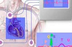
Electrophysiological studies
Last reviewed: 29.06.2025

All iLive content is medically reviewed or fact checked to ensure as much factual accuracy as possible.
We have strict sourcing guidelines and only link to reputable media sites, academic research institutions and, whenever possible, medically peer reviewed studies. Note that the numbers in parentheses ([1], [2], etc.) are clickable links to these studies.
If you feel that any of our content is inaccurate, out-of-date, or otherwise questionable, please select it and press Ctrl + Enter.
Electrophysiologic studies are medical studies that are used to examine the electrical activity of cells and tissues in the human or animal body. These studies are designed to evaluate the heart, nervous system, and other organs in which electrical activity plays an important role. Here are a few types of electrophysiologic studies:
- Electrocardiogram (ECG): This is one of the most common electrophysiologic studies used to examine the electrical activity of the heart. An ECG records the electrical impulses produced by the heart during its contractions and allows the rhythm, frequency and other parameters of the heart to be assessed.
- Electroencephalogram (EEG): This is a study that records the electrical activity of the brain. EEG is used to diagnose various neurological conditions, such as epilepsy, and to study brain activity under different conditions.
- Electromyography (EMG): EMG studies the electrical activity of muscles. This study can help diagnose and monitor muscle and nerve disorders such as neuromuscular diseases.
- Electroneuromyography (ENMG): This is a combination test that uses EMG and nerve stimulation to evaluate nerve and muscle function. It can help identify problems with nerves and muscles.
- Pacing: This is an electrophysiologic study in which electrodes are inserted into the heart to assess its rhythm and conduction. Pacing may be performed to diagnose and treat certain cardiac arrhythmias.
Electrophysiologic studies help physicians better understand the functional status of the body's organs and systems and assist in the diagnosis and treatment of many medical conditions. They are often performed by specialists, such as cardiologists, neurologists and neurophysiologists, using specialized instruments and equipment.
Indications for the procedure
An electrophysiologic study (EPIS) may be recommended to diagnose and monitor a variety of medical conditions and diseases related to the electrical activity of tissues and organs. Indications for EPIS can vary depending on specific clinical symptoms and suspected diagnoses. Below are some common indications for electrophysiologic testing:
- Cardiac arrhythmias: To evaluate the electrical activity of the heart, detect arrhythmias, determine their type, location and cause.
- Heart Block: To diagnose and determine the degree of heart conduction block.
- Glaucoma: To evaluate retinal function and diagnose glaucoma (a disease of the eye associated with increased intraocular pressure).
- Ocularmyasthenia gravis: For the diagnosis of ocular muscle disorders and neuromuscular diseases such as ocular myasthenia gravis.
- Epilepsy: To investigate the electrical activity of the brain and detect epileptic disorders.
- Neuropathies: To evaluate peripheral nerve function and diagnose neuropathies (nerve lesions).
- Myoclonias and tremors: To investigate myoclonias (brief spasmodic muscle movements) and tremors (shaking) in order to diagnose their causes.
- Pediatric Cerebral Palsy: To assess the electrical activity of the brain in children with cerebral palsy.
- Retinal Diseases: For diagnosing and monitoring retinal conditions and evaluating visual function.
- Other neurologic and neuromuscular diseases: For diagnosis and monitoring of other diseases of the nervous and muscular systems.
Technique of the electrophysiological studies
Cardiac electrophysiologic study (EPIS)
This is a medical procedure that is performed to evaluate the electrical activity and rhythm of the heart. This procedure is used to diagnose and treat cardiac arrhythmias, assess heart valve function, and determine the presence and location of electrical conduction pathways in the heart.
Here's how an electrophysiologic study of the heart is done:
- Preparation: The patient may be instructed to take or discontinue certain medications prior to the procedure. The procedure is usually performed in a specialized electrophysiology laboratory (EPL) and the patient will be asked to wear hospital attire prior to the procedure.
- Administrationof local anesthesia: To make the procedure more comfortable for the patient, the area where the intravenous catheter will be inserted is anesthetized locally.
- Intravenous catheter insertion: The doctor inserts thin, flexible catheters through a vein in the groin or neck area and guides them to the heart. These catheters can be used to record the heart's electrical activity and perform electrical tests.
- Electrophysiologic testing: The doctor performs various electrophysiologic tests to evaluate the activity of the heart and determine if arrhythmias are present. These tests may include stimulating the heart, recording electrical activity, and creating electrocardiograms.
- Diagnosis and Treatment: Based on the results of the tests performed, a physician can diagnose arrhythmias, determine their type and location, and decide what treatment measures (such as pacemaker placement or ablation) may be necessary to correct the arrhythmias.
- Completion of the procedure: At the end of the procedure, the catheters are removed and the catheter insertion site is closed.
An electrophysiologic study of the heart is an important diagnostic tool for determining the cause and treatment of cardiac arrhythmias. Doctors who specialize in this procedure are called electrophysiologists.
Electrophysiologic study of the eyes
Generally means performing an electroretinogram (ERG) and/or electrooculogram (EOG), which allow us to study the electrical activity of the eye and its structures, as well as the functional status of the ocular system. Here is a brief description of these two types of electrophysiologic eye examinations:
- Electroretinogram (ERG): This is a study that evaluates the electrical activity of the retina of the eye. The retina is the tissue inside the eye that plays a key role in perceiving light and forming visual images. ERG records the electrical potentials created by the retina in response to light stimuli and can help diagnose various retinal diseases such as retinal degeneration, retinitis, and others.
- Electrooculogram (EOG): This exam evaluates the electrical activity of the eye muscles and eye movement. EOG measures the electrical potentials produced by the eye muscles as they move and fix the gaze. This study can be used to diagnose and monitor eye muscle health and vestibular function.
These electrophysiologic studies of the eye can be useful in diagnosing various diseases and conditions of the ocular system, as well as for evaluating the effectiveness of treatment. They are performed by specialists in ophthalmology and neuro-ophthalmology using specialized instruments and equipment. Doctors may recommend these tests if a patient has symptoms or signs that indicate eye or retinal problems, or to monitor the eye system for certain diseases.
Transesophageal electrophysiologic study (PEIS)
This is a medical procedure that is performed to evaluate the electrical activity of the heart, specifically in the area of the cartilaginous (sterno-cartilaginous) junction. This joint is located between the sternum and the cartilage that connects the sternum to the clavicle.
PEIS is performed to diagnose and treat cardiac arrhythmias, especially those that may be associated with electrical conduction pathways through the cartilage articulation. This procedure may be recommended if conventional electrocardiograms (ECGs) and cardiac electrophysiologic studies performed through intravenously inserted catheters do not provide sufficient information.
This is how a transesophageal electrophysiologic study is performed:
- Preparation: Preparation for CEIS involves the same steps as preparation for a normal electrophysiologic cardiac study. This may include taking or stopping medications, as well as making sure to check with your doctor.
- Catheter insertion: The doctor inserts a thin, flexible catheter through a vein in the groin or neck area and guides it to the cartilaginous articulation.
- Electrophysiologic Testing: After insertion of the catheter, the physician performs various electrophysiologic tests to assess the electrical activity of the heart in the cartilage articulation area.
- Diagnosis and Treatment: The doctor uses the test results to diagnose arrhythmias, determine their type and location, and decide on treatment options, such as pacemaking or ablation, if necessary.
- Completion of the procedure: At the end of the procedure, the catheter is removed and the insertion site is closed.
PEIS is an important tool for investigating and treating arrhythmias associated with cartilage articulation and can help physicians accurately diagnose and treat these conditions. This procedure is performed by specialized medical teams including electrophysiologists and cardiologists.
Intracardiac electrophysiologic study (IVEPI)
Also known as an electrophysiologic cardiac examination (EPIS), is a procedure performed in cardiology to evaluate the electrical activity of the heart and to determine the causes and treatment of various cardiac arrhythmias and heart rhythm disorders. This study is usually performed in a specialized clinic or cardiac center and requires special equipment and trained medical personnel.
Here's how the WSEPI goes:
- Patient preparation: The patient may require some preparation before the procedure, including fasting before the test (e.g., not eating or drinking for several hours before the procedure) and consenting to the procedure.
- Monitoring set-up: The patient may be fitted with electrodes (electrocardiographic or ECG electrodes) on the chest where the electrical activity of the heart will be monitored during the study.
- Local anesthesia: Under local anesthesia (or sometimes general anesthesia), medical personnel insert catheters (thin, flexible tubes) through blood vessels (usually a vein in the groin or arm) and guide them to the heart.
- Measuring electrical activity: Specialists use these catheters to record the electrical signals generated by the heart. This allows them to assess the electrical activity of different parts of the heart and detect abnormalities.
- Inducing arrhythmias: During VSEPI, cardiac stimulation can be performed to induce arrhythmias and determine their causes and mechanisms. This can help specialists determine the best way to treat the arrhythmia.
- Treatment: In some cases, if arrhythmias or other cardiac abnormalities are detected, therapeutic manipulations such as ablation (removal or isolation of abnormal areas of heart tissue) may be performed during VSEPI.
-
Completion of the procedure: Once the study is complete, all catheters are removed and the catheter insertion site is closed. The patient may need some time to recover from the procedure.
VSEPI is an important tool for the diagnosis and treatment of cardiac arrhythmias and heart rhythm disorders. It may be recommended by a physician when other diagnostic methods fail to fully understand the cause or mechanism of an arrhythmia.
Electrophysiological methods of hearing research
Hearing research using electrophysiologic techniques assesses the functional status of the ear and auditory system by measuring electrical signals and nervous system activity in response to acoustic stimuli. These methods can be useful in the diagnosis of auditory disorders and hearing impairment. Some electrophysiologic methods for the study of hearing are presented below:
- Evoked potential audiometry (ABR/BERA): This is one of the most common electrophysiologic methods. The patient is provided with an earpiece through which a series of click or tone sound pulses are delivered. Electrodes placed on the patient's scalp and ear then record the evoked potentials that occur in the nervous system in response to the acoustic stimuli. These potentials allow the functional status of the auditory nerves and auditory pathways to be assessed.
- Auditory stimulation of the inner ear (ECochG): This method allows the electrophysiological activity of the inner ear to be studied and the function of auditory organs such as the cochlea and vestibular apparatus to be assessed. Electrodes are inserted inside the ear drum and can record responses to sound and electrical stimuli.
- Bone conduction auditorystimulation using osteophones (BCER): This method assesses auditory function by transmitting sound waves directly through the bone conduction of the skull. Electrodes are placed on the scalp and stimuli are sent using vibrations. This method is useful in diagnosing auditory disorders associated with outer and middle ear disorders.
- Evoked midbrain potentials (MMN, P300): These electrophysiological techniques can be used to study higher auditory functions such as the recognition and processing of sound information in the brain. They can be useful in evaluating cognitive aspects of hearing and detecting neurological disorders.
These electrophysiologic techniques can be useful in the diagnosis of various auditory disorders, including auditory neuritis, hearing impairment in newborns and infants, and evaluating the effectiveness of hearing prostheses and implants. These studies are performed by specialists in audiology and orthopedics.
Electrophysiologic study of the optic nerve
It is not usually a standardized examination procedure. Instead, various methods and examinations such as ophthalmoscopy, electroretinography (ERG), electro-oculogram (EOG), and Visual Evoked Potentials (VEP) are used to assess optic nerve function and eye health.
Here's a brief description of some of these methods:
- Ophthalmoscopy: This method allows the doctor to examine the eye fundus and optic nerve with a specialized instrument called an ophthalmoscope. It is used to detect changes in the eye such as optic nerve swelling, degeneration, or other abnormalities.
- Electroretinography (ERG): ERG is an electrophysiologic study that records the electrical activity of the retina in response to light stimuli. It helps in the assessment of retinal function and early diagnosis of a number of eye diseases.
- Electrooculogram (EOG): EOG is a method for studying eyeball movements and ophthalmic muscle function. It can be useful in diagnosing some neurological or eye disorders.
- Visual Evoked Potential (VEP) study: VEP is an electrophysiologic study that records the electrical activity of the brain in response to visual stimuli. It can be used to assess optic nerve function and diagnose neurological or ocular disorders.
Electrophysiological methods of central nervous system research
Allow you to study the electrical activity and functional characteristics of the brain and spinal cord. These techniques are important tools in neurophysiology and can help in the diagnosis of various neurological conditions and the scientific study of CNS functions. Here are some of the most common electrophysiologic methods used to study the CNS:
- Electroencephalogram (EEG): An EEG records the electrical activity of the brain using electrodes placed on the scalp. This study examines electrical patterns of brain activity and can be used to diagnose epilepsy, assess the functional state of the brain in various diseases, and in sleep and neurophysiology research.
- Electromyography (EMG): EMG evaluates the electrical activity of muscles using electrodes inserted into the muscles. This study can help diagnose muscle and nerve disorders such as neuromuscular diseases.
- Electroneuromyography (ENMG): ENMG is a combination test that combines EMG and nerve stimulation to evaluate nerve and muscle function. It can help identify problems with nerves and muscles.
- Visually Evoked Potentials (VEPs): This method examines the electrical activity of the brain occurring in response to visual stimuli such as light flashes or patterns. VEPs can be used to diagnose visual diseases and assess visual function.
- Somatosensory Evoked Potentials (SVPs): This method examines the electrical activity of the brain associated with somatic (bodily) sensations, such as skin sensations or limb positions. SVPs are used in clinical practice to diagnose neurological disorders.
Electrophysiological methods of oral receptor research
Used to study the electrical signals generated by receptors in the mouth when they interact with different chemicals, tastes and odors. These techniques can help us understand what signals are sent to the brain in response to stimulation of taste and smell receptors, and how this affects our perception of food and flavors. Here are some electrophysiological techniques that can be used in studies of oral receptors:
- Electroglottography (EGG): This method is used to study the movements and electrical activity of the muscles of the larynx and pharynx when swallowing and perceiving taste stimuli. It can help assess responses to different food flavors and textures.
- Electroencephalography (EEG): EEG measures the electrical activity of the brain. This method can be used to study the activation of brain regions associated with the perception of taste and smell stimuli.
- Electromyography (EMG): EMG measures the electrical activity of muscles. It can be used to assess the activity of the chewing muscles and other muscles associated with the eating process.
- Intracellular recording of receptor action potential: This method records electrical signals directly from receptor cells in the oral cavity. It can be useful for studying receptor responses to different chemicals and taste stimulants.
- Receptor Field Potential: This method measures changes in the electrical potential around oral receptors in response to stimulation. It can be used to study the perception of tastes and odors.
Electrophysiological methods for studying oral receptors provide a better understanding of how the sensory organs in the oral cavity interact with different stimuli and how this information is transmitted to the brain. These techniques can be useful in studies of taste and olfactory receptors, as well as in the development of new products and treatments related to oral sensitivity.
Contraindications to the procedure
Electrophysiologic testing methods such as EEG, EMG, GDP and others are generally safe, but they may also have some contraindications and limitations. Contraindications may vary depending on the specific study method and clinical situation. Some common contraindications are summarized below:
- Allergic reaction to electrodes or gel: In rare cases, an allergic reaction to materials used in electrophysiologic studies, such as electrodes or gel, may occur. Patients with known allergies to these components may be at risk.
- Infectious Diseases: Electrophysiologic studies that involve the insertion of electrodes or needles into the body may be contraindicated in active infectious diseases such as purulent skin infections.
- Recent surgery or trauma: Some electrophysiologic studies may be limited or contraindicated in the case of recent surgery or trauma, especially if it is related to the area to be studied.
- Epilepsy and neurologic disorders: Some patients with epilepsy or other severe neurologic disorders may have limitations on electrophysiologic studies.
- Uncooperative: Children or patients who are unable to cooperate and follow instructions during the study may have limitations in electrophysiologic procedures.
Complications after the procedure
Cardiac electrophysiologic study (EPIS) and electrophysiologic study of other organs and systems can be relatively safe procedures, but like any medical procedure, they can come with certain risks and complications. Here are some of the possible complications after an EPIS:
- Bleeding or hematoma: Bleeding or hematoma formation may occur at the catheter insertion site. This complication may require medical attention.
- Infection: Any intravenous intervention, including catheter insertion, can be a source of infection. Adherence to strict sterile conditions is important to prevent this complication.
- Reaction to the contrast agent: In some cases, there may be an allergic reaction to the contrast agent used in the procedure.
- Arrhythmias: The study itself may occasionally cause temporary cardiac arrhythmias that may require medical supervision.
- Embolism: An indwelling catheter can be a source of blood clots or emboli (blood vessel blockages) that can cause serious complications.
- Vascular or tissue damage: Improper catheter insertion or maneuvering can cause damage to blood vessels, heart muscle, or other tissues.
- Pain or discomfort: You may experience pain, discomfort, or discomfort in the catheter insertion area after the procedure.
- Allergic reactions to anesthetics: If local anesthesia is used, allergic reactions to anesthetics may occur.
- Possible complications within an organ: Depending on the specific purpose of the study, there may be specific complications associated with assessing the function of that organ. For example, arrhythmias or perforation of the heart wall may occur during cardiac EPIS.
Care after the procedure
Care after electrophysiology procedures depends on the specific study and the instructions provided by the medical professional or technician performing the study. However, there are general guidelines that may help you after these procedures:
- Talk to your doctor: After the procedure is complete, discuss the results of the test with your doctor. Your doctor can provide important information about what was found and what further steps are needed.
- Follow recommendations: Your doctor or health care professional can give you recommendations for actions you should take after the procedure. This may include instructions on taking medication, diet, or other aspects of your health care.
- Rest and recovery: Some procedures may be tiring or may cause discomfort. Rest and allow your body to recover.
- Care of the procedure site: If you have had electrodes or needles placed, follow your doctor's instructions regarding the care of these sites. This may include keeping the skin dry and clean and avoiding painful movements.
- Restrictions: You may need to follow activity or dietary restrictions in some cases. Make sure you understand and follow these restrictions.
- Keep an eye out for possible complications: If you experience any unusual symptoms or complications after the procedure, contact your doctor immediately. This may include pain, swelling, bleeding, or other changes you notice.
- Maintain your follow-up regimen: If you have follow-up care or additional procedures, follow instructions and come to your doctor's appointments.