Medical expert of the article

New publications
Electroencephalography in epilepsy
Last reviewed: 04.07.2025

All iLive content is medically reviewed or fact checked to ensure as much factual accuracy as possible.
We have strict sourcing guidelines and only link to reputable media sites, academic research institutions and, whenever possible, medically peer reviewed studies. Note that the numbers in parentheses ([1], [2], etc.) are clickable links to these studies.
If you feel that any of our content is inaccurate, out-of-date, or otherwise questionable, please select it and press Ctrl + Enter.
Epilepsy is a disease manifested by two or more epileptic seizures (fits). An epileptic seizure is a short, usually unprovoked, stereotypical disorder of consciousness, behavior, emotions, motor or sensory functions, which even by clinical manifestations can be associated with the discharge of an excessive number of neurons in the cerebral cortex. The definition of an epileptic seizure through the concept of neuron discharge determines the most important significance of EEG in epileptology. Clarification of the form of epilepsy (more than 50 variants) includes a mandatory component description of the EEG pattern characteristic of this form. The value of EEG is determined by the fact that epileptic discharges, and therefore epileptiform activity, are observed on EEG outside of an epileptic seizure.
Reliable signs of epilepsy are discharges of epileptiform activity and patterns of epileptic seizures. In addition, high-amplitude (more than 100-150 μV) bursts of alpha, delta, and theta activity are characteristic, but they cannot be considered evidence of epilepsy in themselves and are assessed in the context of the clinical picture. In addition to the diagnosis of epilepsy, EEG plays an important role in determining the form of the epileptic disease, which determines the prognosis and choice of drug. EEG allows you to select a drug dose based on the assessment of the reduction in epileptiform activity and predict side effects based on the appearance of additional pathological activity.
To detect epileptiform activity on the EEG, rhythmic light stimulation (mainly in photogenic seizures), hyperventilation or other effects are used, based on information about the factors that provoke seizures. Long-term recording, especially during sleep, helps to detect epileptiform discharges and patterns of epileptic seizures. Sleep deprivation helps to provoke epileptiform discharges on the EEG or the seizure itself. Epileptiform activity confirms the diagnosis of epilepsy, but is also possible in other conditions, while in some patients with epilepsy it cannot be recorded.
 [
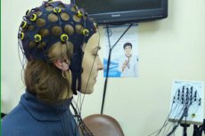
[ Long-term recording of electroencephalogram and EEG video monitoring
Like epileptic seizures, epileptiform activity on the EEG is not recorded constantly. In some forms of epileptic disorders, it is observed only during sleep, sometimes provoked by certain life situations or forms of patient activity. Consequently, the reliability of epilepsy diagnostics directly depends on the possibility of long-term EEG recording under conditions of sufficiently free behavior of the subject. For this purpose, special portable systems for long-term (12-24 hours or more) EEG recording under conditions close to normal life activity have been developed. The recording system consists of an elastic cap with specially designed electrodes built into it, allowing for long-term high-quality EEG recording. The recorded electrical activity of the brain is amplified, digitized and recorded on flash cards by a recorder the size of a cigarette case, which fits in a convenient bag on the patient. The patient can perform normal household activities. After the recording is completed, the information from the flash card in the laboratory is transferred to a computer system for recording, viewing, analyzing, storing and printing electroencephalographic data and is processed as a regular EEG. The most reliable information is provided by EEG video monitoring - simultaneous registration of the EEG and video recording of the patient during an attack. The use of these methods is required in the diagnosis of epilepsy when routine EEG does not reveal epileptiform activity, as well as in determining the form of epilepsy and the type of epileptic seizure, for the differential diagnosis of epileptic and non-epileptic seizures, clarification of the goals of surgery during surgical treatment, diagnosis of epileptic non-paroxysmal disorders associated with epileptiform activity during sleep, control of the correctness of the choice and dose of the drug, side effects of therapy, reliability of remission.
Electroencephalogram characteristics in the most common forms of epilepsy and epileptic syndromes
- Benign epilepsy of childhood with centrotemporal spikes (benign rolandic epilepsy).
- Outside the seizure: focal spikes, sharp waves and/or spike-slow wave complexes in one hemisphere (40-50%) or in both hemispheres with unilateral predominance in the central and middle temporal leads, forming antiphases over the rolandic and temporal areas. Sometimes epileptiform activity is absent during wakefulness, but appears during sleep.
- During an attack: focal epileptic discharge in the central and middle temporal leads in the form of high-amplitude spikes and sharp waves, combined with slow waves, with possible spread beyond the initial localization.
- Benign occipital epilepsy of childhood with early onset (Panayotopoulos form).
- Outside of an attack: 90% of patients show mainly multifocal high- or low-amplitude sharp-slow wave complexes, often bilaterally synchronous generalized discharges. In two-thirds of cases, occipital spikes are observed, in a third of cases - extraoccipital. Complexes occur in series when the eyes are closed. Blocking of epileptiform activity by opening the eyes is noted. Epileptiform activity on the EEG and sometimes seizures are provoked by photostimulation.
- During an attack: an epileptic discharge in the form of high-amplitude spikes and sharp waves, combined with slow waves, in one or both occipital and posterior parietal leads, usually with spread beyond the initial localization.
- Idiopathic generalized epilepsies. EEG patterns characteristic of childhood and juvenile idiopathic epilepsy with absences, as well as of juvenile idiopathic myoclonic epilepsy. EEG characteristics in primary generalized idiopathic epilepsy with generalized tonic-clonic seizures are as follows.
- Outside of an attack: sometimes within normal limits, but usually with moderate or marked changes with delta, theta waves, bursts of bilaterally synchronous or asymmetric spike-slow wave complexes, spikes, sharp waves.
- During an attack: a generalized discharge in the form of rhythmic activity of 10 Hz, gradually increasing in amplitude and decreasing in frequency in the clonic phase, sharp waves of 8-16 Hz, spike-slow wave and polyspike-slow wave complexes, groups of high-amplitude delta and theta waves, irregular, asymmetrical, in the tonic phase delta and theta activity, sometimes ending in periods of no activity or low-amplitude slow activity.
- Symptomatic focal epilepsies: the characteristic epileptiform focal discharges are observed less regularly than in idiopathic ones. Even seizures may not present with typical epileptiform activity, but with bursts of slow waves or even desynchronization and seizure-related flattening of the EEG.
- In limbic (hippocampal) temporal epilepsies, there may be no changes in the interictal period. Usually, focal sharp-slow wave complexes are observed in the temporal leads, sometimes bilaterally synchronous with unilateral amplitude predominance. During an attack, there are bursts of high-amplitude rhythmic "steep" slow waves, or sharp waves, or sharp-slow wave complexes in the temporal leads spreading to the frontal and posterior leads. At the beginning (sometimes during) the seizure, unilateral flattening of the EEG may be observed. In lateral temporal epilepsies with auditory and, less frequently, visual illusions, hallucinations and dreamlike states, speech and orientation disorders, epileptiform activity on the EEG is observed more often. Discharges are localized in the middle and posterior temporal leads.
- In non-convulsive temporal seizures occurring as automatisms, a picture of epileptic discharge is possible in the form of rhythmic primary or secondary generalized high-amplitude delta activity without acute phenomena, and in rare cases - in the form of diffuse desynchronization, manifested by polymorphic activity with an amplitude of less than 25 μV.
- In frontal lobe epilepsies, EEG does not reveal focal pathology in the interictal period in two thirds of cases. In the presence of epileptiform oscillations, they are recorded in the frontal leads on one or both sides, bilaterally synchronous spike-slow wave complexes are observed, often with lateral predominance in the frontal areas. During a seizure, bilaterally synchronous spike-slow wave discharges or high-amplitude regular delta or theta waves can be observed, mainly in the frontal and/or temporal leads, sometimes sudden diffuse desynchronization. In orbitofrontal foci, three-dimensional localization reveals the corresponding location of the sources of the initial sharp waves of the epileptic seizure pattern.
- Epileptic encephalopathies. A new diagnostic rubric has been introduced into the proposals of the Terminology and Classification Commission of the International League Against Epilepsy, which includes a wide range of severe epileptic disorders: epileptic encephalopathies. These are permanent disorders of brain function caused by epileptic discharges that manifest themselves on the EEG as epileptiform activity, and clinically as various long-term mental, behavioral, neuropsychological, and neurological disorders. These include West infantile spasm syndrome, Lennox-Gastaut syndrome, other severe "catastrophic" infantile syndromes, as well as a wide range of mental and behavioral disorders that often occur without epileptic seizures. Diagnosis of epileptic encephalopathy is possible only with the help of EEG, since in the absence of seizures only it can establish the epileptic nature of the disease, and in the presence of seizures, it can clarify the affiliation of the disease specifically to epileptic encephalopathy. Below are data on EEG changes in the main forms of epileptic encephalopathy.
- West's infantile spasms syndrome.
- Outside of an attack: hypsarrhythmia, i.e. continuous generalized high-amplitude slow activity and sharp waves, spikes, spike-slow wave complexes. There may be local pathological changes or persistent asymmetry of activity.
- During an attack: the lightning-fast initial phase of the spasm corresponds to generalized spikes and sharp waves, tonic convulsions - generalized spikes, increasing in amplitude towards the end of the attack (beta activity). Sometimes the attack is manifested by a suddenly arising and ending desynchronization (decrease in amplitude) of the current epileptiform high-amplitude activity.
- Lennox-Gastaut syndrome.
- Outside of an attack: continuous generalized high-amplitude slow and hypersynchronous activity with sharp waves, spike-slow wave complexes (200-600 μV), focal and multifocal disturbances corresponding to the picture of hypsarrhythmia.
- During an attack: generalized spikes and sharp waves, spike-slow wave complexes. During myoclonic-astatic seizures - spike-slow wave complexes. Sometimes desynchronization is noted against the background of high-amplitude activity. During tonic seizures - generalized high-amplitude (>50 μV) sharp beta activity.
- Early infantile epileptic encephalopathy with burst-suppression pattern on EEG (Ohtahara syndrome).
- Outside of an attack: generalized burst-suppression activity - 3-10 second periods of high-amplitude 9, 5 activity with irregular asymmetrical polyspike-slow wave, sharp-slow wave complexes of 1-3 Hz, interrupted by periods of low-amplitude (<40 μV) polymorphic activity, or hypsarrhythmia - generalized 8 and 9 activity with spikes, sharp waves, spike-slow wave, polyspike-slow wave, sharp-slow wave complexes with an amplitude of more than 200 μV.
- During an attack: an increase in the amplitude and number of spikes, sharp waves, spike-slow wave complexes, polyspike-slow wave, sharp-slow wave with an amplitude of more than 300 μV, or flattening of the background recording.
- Epileptic encephalopathies manifested mainly by behavioral, mental and cognitive disorders. These forms include Landau-Kleffner epileptic aphasia, epilepsy with constant spike-slow wave complexes in slow-wave sleep, frontal lobe epileptic syndrome, acquired epileptic syndrome of right hemisphere developmental disorder, and others. Their main feature and one of the main diagnostic criteria is gross epileptiform activity corresponding in type and localization to the nature of the impaired brain function. In general developmental disorders such as autism, bilaterally synchronous discharges characteristic of absences may be observed, in aphasia - discharges in the temporal leads, etc.