Medical expert of the article

New publications
Ear otoscopy: what is it?
Last reviewed: 03.07.2025

All iLive content is medically reviewed or fact checked to ensure as much factual accuracy as possible.
We have strict sourcing guidelines and only link to reputable media sites, academic research institutions and, whenever possible, medically peer reviewed studies. Note that the numbers in parentheses ([1], [2], etc.) are clickable links to these studies.
If you feel that any of our content is inaccurate, out-of-date, or otherwise questionable, please select it and press Ctrl + Enter.
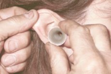
In otolaryngology, a special diagnostic procedure called otoscopy is performed to examine the external auditory canal and examine the eardrum.
Indications for the procedure
Otoscopy is performed during routine medical examinations and also on patients complaining of ear pain, ringing or noise in the ears, discomfort or itching in the external auditory canal, otorrhea (discharge from one or both ears) and hearing loss.
In addition, otoscopy is used to perform appropriate medical procedures as prescribed: foreign bodies are removed from the ear canal and accumulated exudate or pus is removed from the middle ear cavity (located behind the eardrum) by puncturing the eardrum (paracentesis) or opening it (tympanotomy or myringotomy).
Otoscopy of the ear and eardrum (membrana tympani), which separates the external auditory canal from the middle ear (auris media), allows one to assess the condition of visible anatomical structures and diagnose inflammation of the auditory canal and diseases of the middle ear, including acute otitis media and its complications; purulent otitis, including chronic.
Using visualization, perforation of the eardrum of any etiology, as well as otomycosis (fungal infection of the ear, fungal otitis) can be detected.
Preparation
Earwax accumulation - a wax plug during otoscopy prevents it from being performed, so preparation for the procedure involves the doctor removing the wax and cleaning the external auditory canal from skin scales (keratin debris), crusts, etc.
If the procedure is scheduled in advance, it is recommended to postpone washing the ears or using ear drops.
Technique otoscopies
The technique for examining the external auditory canal and eardrum has long been developed, but the types of otoscopy may determine some of its variations.
The classic type of otoscopy is with an ear funnel (ear mirror), a head reflector (a round mirror with a hole in the center) and an electric lamp, the light of which is reflected by the reflector. Nowadays, medical head lights with batteries or accumulators are used. [ 1 ]
A more modern examination of the ear is performed with a special monocular otoscope (consisting of a handle and a head), on the front end of which there is an attachment for disposable plastic ear funnels, and in the head there is an independent light source and a lens with three-fold magnification.
Video otoscopy or endoscopic otoscopy – using a digital optical otoscope (with a light source and a miniature video camera) inserted into the external auditory canal – allows the doctor to obtain a clear image on a color monitor.
Pneumatic otoscopy is used to determine the mobility of the intact tympanic membrane under induced pressure changes, which are provided by a pneumatic balloon connected to the otoscope. Stability of the tympanic membrane in response to pressure can be caused by fluid in the middle ear, and this type of otoscopy is considered the mainstay in the diagnosis of otitis media with effusion. A pneumatic otoscope can also be useful in differentiating the degree of perforation of the tympanic membrane.[ 2 ]
Visualization of the ear canal and eardrum using a binocular microscope (with the patient lying on his back with his head tilted) is called microscopic otoscopy or otomicroscopy. It provides a wider field of view and 40x magnification of anatomical structures.
Before the otoscopic examination, an experienced doctor will check the condition of the facial (VII cranial) nerve passing through the middle ear: the patient is asked to smile, frown, puff out his cheeks, and raise his eyebrows with his eyes closed. Then a physical examination of the auricle (with its palpation) and the area behind the ear is performed.
The sequence of actions – the otoscopy algorithm – includes:
- choosing an ear funnel that is the right size for the patient's ear canal;
- insertion of a funnel with straightening of the external auditory canal, for which the auricle is pulled back and up in adult patients, and back and down in children. Only after this is the ear speculum carefully inserted into the auditory canal, and the doctor examines it;
- slowly moving the otoscope funnel into the canal until the eardrum becomes visible, the condition of which is assessed taking into account the color, the presence of a bulge and perforation. The doctor also observes the so-called landmarks of the eardrum: the three-layered stretched part (pars tensa), the two-layered relaxed part (pars flaccida) and the handle of the malleus (malleus) - the largest auditory ossicle of the middle ear, adjacent to the tympanic membrane;
- slowly removing the funnel from the ear canal.
Otoscopic signs of otitis and other diseases
What can a doctor see with otoscopy? If there is no otitis or other ear diseases, normal otoscopy means visualization of the normal eardrum at the end of the external auditory canal - a translucent pale gray (whitish) membrane of an oval shape (in childhood it is round).
In acute otitis externa, the skin of the ear canal is painful and swollen, and visualization of the eardrum may not be possible.
In the early stages of acute otitis media, the eardrum changes depending on the stage of the disease. At first, it is pink, retracted, with dilated peripheral vessels. As the inflammatory process progresses, the eardrum swells, becomes bright red; it may perforate with the release of pus into the external auditory canal. [ 3 ]
In exudative otitis media, the eardrum is retracted and immobile, and due to serous effusion it becomes yellowish.
Read also – Diagnosis of acute otitis media
Otoscopy in chronic purulent otitis media can detect both of its forms: mesotympanitis and epitympanitis. The main otoscopic signs of mesotympanitis are through perforation of various shapes and sizes of the stretched part of the eardrum with its redness and edema and/or granulation along the edges of the opening. And epitympanitis is distinguished by a violation of the integrity of the eardrum along the edges of its unstretched part.
Otoscopy in otomycosis reveals fluffy-looking white or cream-colored particles. If the infection is caused by Aspergillus niger, tiny grayish-black mycelial outgrowths may be seen.
The growth of new spongy bone tissue around the supporting plate of the stapes of the middle ear in the area of the oval window - otosclerosis - is difficult to diagnose during an otoscopic examination, since the pathological process develops in the tympanic cavity. And the otologist can observe a change in the color of the eardrum and its thinning, as well as redness of the mucous membrane covering the tympanic cavity (which is visible through the eardrum).
Mastoiditis is an inflammation of the mastoid process (processus mastoideus) of the temporal bone of the skull located behind the ear, the tympanic and squamous parts of which limit the auditory opening and the external auditory canal on three sides. During otoscopy, deformation of part of the wall of the external auditory canal formed by the tympanic and squamous bones is visualized. The main method of instrumental diagnostics of this disease is MRI. [ 4 ]
Contraindications to the procedure
Otoscopy is performed on children of any age and adults. In addition to the technical complexity of anatomical anomalies of the ears and stenosis of the external auditory canal, contraindications to its implementation include severe swelling of the auditory canal and the presence of strong bloody, serous or purulent discharge from the auditory opening. [ 5 ]
Complications after the procedure
Insertion of an ear speculum into the external auditory canal can cause a reflex dilation of the blood vessels supplying the eardrum, leading to temporary hyperemia in the ear.
Given the frequent use of ear specula and otoscopes, they represent a potential source of pathogenic microorganisms. And the consequence after the procedure - without proper disinfection of the instruments - may be the development of an infection.
When the otoscope is inserted too deeply into the ear canal, or the patient has a very thin eardrum, there is a small risk of damaging the eardrum.
Patients with perforation of the eardrum or rupture of one of the membranes separating the middle and inner ear (perilymph fistula) may have complications after pneumatic otoscopy in the form of dizziness, imbalance, nystagmus, nausea and vomiting.
Reviews
Feedback from ENT doctors confirms the value of information about a possible middle ear disease obtained through direct observation of the eardrum and external auditory canal through an otoscope, which allows for an accurate determination of the cause of the patient's complaints.