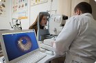
Confocal scanning laser ophthalmoscopy is a method for forming and analyzing a three-dimensional topographic image of the optic nerve head in real time.
With scanning laser polarimetry (SLP), the peripapillary thickness of the optic nerve is determined by measuring the total birefringence of the fundus.
Glaucoma parameters are measured by assessing the optic disc excavation, SNV defects, and possibly their thickness ratio at the macula. These parameters are reliable indicators of glaucoma and its progression.
Electroretinography objectively establishes retinal dysfunction. With multifocal electroretinography, focal responses are obtained from a large number of retinal areas and topographic maps of areas with impaired function are constructed.
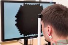
Dual frequency perimetry (DFP) technology (Welch Allyn, Skaneateles, NY, and Humphrey Systems, Dublin, CA) is used for effective early visual field assessment and detection of glaucomatous visual field changes.
Glaucoma is a common cause of blindness in all countries and can develop in any age group, but is especially common after 40 years of age. Increased intraocular pressure is the most important causal risk factor for glaucoma, but high intraocular pressure is not necessary for the development of glaucomatous damage.
In ultrasound biomicroscopy (UBM) of the anterior segment, high-frequency transducers (50 MHz) are used to obtain high-resolution images (approximately 50 µm), allowing in vivo imaging of the anterior segment of the eye (penetration depth - 5 mm)
It has been established that the goal of glaucoma treatment is to prevent further development of symptomatic vision loss with maximum reduction of side effects or complications after surgical interventions.