Medical expert of the article
New publications
Classification of breast cancer
Last reviewed: 04.07.2025

All iLive content is medically reviewed or fact checked to ensure as much factual accuracy as possible.
We have strict sourcing guidelines and only link to reputable media sites, academic research institutions and, whenever possible, medically peer reviewed studies. Note that the numbers in parentheses ([1], [2], etc.) are clickable links to these studies.
If you feel that any of our content is inaccurate, out-of-date, or otherwise questionable, please select it and press Ctrl + Enter.

The diversity of morphological features, variants of clinical manifestations and reactions to therapeutic effects give every reason to define breast cancer as a heterogeneous disease. Therefore, today there is not one classification of breast cancer, but several. And each of them is based on its own principles.
TNM classification of breast cancer
The stages of breast cancer are determined by the TNM Classification of Malignant Tumors, adopted by WHO for all malignant neoplasms. For oncological mammology, based on the recommendations of leading specialists, it has been adapted with the introduction of details.
The TNM classification of breast cancer measures the anatomical grade of the tumor based on its size, spread to lymph nodes in the armpits, neck, and chest, and the presence of metastases. This international classification of breast cancer is adopted by the International Association for Breast Cancer and the European Society for Medical Oncology (EUSOMA).
According to the TNM classification, breast cancer has the following stages:
- T0 – signs of breast cancer are not detected (not proven).
- Tis (tumor in situ) designation refers to carcinomas and is deciphered as follows: abnormal cells are found in situ (no invasion), localization is limited to ducts (DCIS) or lobules (LCIS) of the mammary gland. There is also Tis Paget, that is, Paget's disease, affecting the tissues of the nipple and areola of the breast.
- T1 – tumor diameter at its widest point is 20 mm or less:
- T1a – tumor diameter > 1 mm, but < 5 mm;
- T1b – tumor diameter is greater than 5 mm but less than 10 mm;
- T1c – tumor diameter >10 mm but ≤ 20 mm.
- T2 – tumor diameter > 20 mm, but < 50 mm.
- T3 – the tumor diameter exceeds 50 mm.
- T4 – the tumor is any size and has spread: to the chest (T4a), to the skin (T4b), to the chest and skin (T4c), inflammatory breast cancer (T4d).

Lymph node indicators:
- NX – lymph nodes cannot be assessed.
- N0 – cancer was not found in the lymph nodes.
- N0 (+) – small areas of “isolated” tumor cells (less than 0.2 mm) are found in the axillary lymph nodes.
- N1mic – areas of tumor cells in the axillary lymph nodes larger than 0.2 mm but smaller than 2 mm (may only be visible under a microscope and are often called micrometastases).
- N1 – cancer has spread to 1-2-3 axillary lymph nodes (or the same number of intrathoracic lymph nodes), maximum size 2 mm.
- N2 – cancer has spread to 4-9 lymph nodes: only to the axillary (N2a), only to the internal mammary (N2b).
- N3 – the cancer has spread to 10 or more lymph nodes: to the lymph nodes under the arm, or under the collarbone, or above the collarbone (N3a); to the internal mammary or axillary nodes (N3b); the supraclavicular lymph nodes are affected (N3c).
Indicators for distant metastases:
- M0 – no metastases;
- M0 (+) – there are no clinical or radiological signs of distant metastases, but tumor cells are detected in the blood or bone marrow, or in other lymph nodes;
- M1 – metastases in other organs are detected.
Histological classification of breast cancer
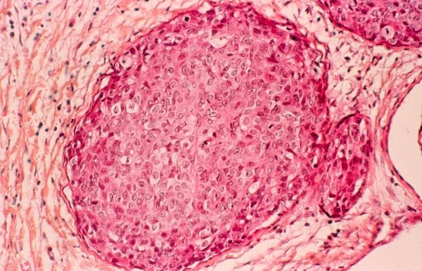
The current histopathological classification of breast cancer is based on the morphological features of neoplasms, which are studied during histological examination of tumor tissue samples – biopsies.
In the current version, approved by the WHO in 2003 and accepted worldwide, this classification includes about two dozen major types of tumors and almost as many less significant (rare) subtypes.
The following main histotypes of breast cancer are distinguished:
- noninvasive (noninfiltrating) cancer: intraductal (ductal) carcinoma; lobular carcinoma (LCIS);
- invasive (infiltrating) cancer: ductal (intraductal) or lobular cancer.
According to statistics from the European Society for Medical Oncology (ESMO), these types account for 80% of clinical cases of malignant breast tumors. In other cases, less common types of breast cancer are diagnosed, in particular: medullary (soft tissue cancer); tubular (cancer cells form tubular structures); mucinous or colloid (with mucus); metaplastic (squamous cell, glandular-squamous cell, adenoid cystic, mycoepidermoid); papillary, micropapillary); Paget's disease (tumor of the nipple and areola), etc.
Based on the standard histological examination protocol, the level of differentiation (distinction) of normal and tumor cells is determined, and thus the histological classification of breast cancer allows us to establish the degree of tumor malignancy (this is not the same as cancer stages). This parameter is very important, since the level of histopathological differentiation of neoplastic tissue gives an idea of the potential for its invasive growth.
Depending on the number of deviations in the cell structure, degrees are distinguished (Grade):
- GX – tissue discrimination level cannot be assessed;
- G1 – the tumor is highly differentiated (low grade), that is, the tumor cells and organization of the tumor tissue are close to normal;
- G2 – moderately differentiated (middle grade);
- G3 – low differentiated (high grade);
- G4 – undifferentiated (high grade).
Grades G3 and G4 indicate a significant predominance of atypical cells; such tumors grow rapidly and their rate of spread is higher than that of tumors with differentiation at the G1 and G2 level.

The main drawbacks of this classification, according to experts, are the limited ability to more accurately reflect the heterogeneity of breast cancer, since one group included tumors with completely different biological and clinical profiles. As a result, the histological classification of breast cancer has minimal prognostic value.
Immunohistochemical classification of breast cancer
Thanks to the use of new molecular tumor markers – the expression of tumor cell receptors for estrogen (ER) and progesterone (PgR) and the status of HER2 (transmembrane protein receptor of the epidermal growth factor EGFR, stimulating cell growth) – a new international classification of breast cancer has emerged, which has proven prognostic value and allows more accurate determination of therapy methods.
Based on the state of estrogen and progesterone receptors, the activation of which leads to changes in cells and tumor growth, the immunohistochemical classification of breast cancer distinguishes between hormone-positive tumors (ER+, PgR+) and hormone-negative (ER-, PgR-). Also, the status of EGFR receptors can be positive (HER2+) or negative (HER2-), which fundamentally affects the treatment tactics.
Hormone-positive breast cancer responds to hormone therapy with drugs that lower estrogen levels or block its receptors. These tumors tend to grow more slowly than hormone-negative ones.
Mammologists note that patients with this type of neoplasm (which often occurs after menopause and affects the tissues lining the ducts) have a better prognosis in the short term, but cancer with ER+ and PgR+ can sometimes recur after many years.
Hormone-negative tumors are much more often diagnosed in women who have not yet gone through menopause; these neoplasms are not treated with hormonal drugs and grow faster than hormone-positive cancers.
In addition, the immunohistochemical classification of breast cancer distinguishes triple positive cancer (ER+, PgR+ and HER2+), which can be treated with hormonal agents and drugs with monoclonal antibodies designed to suppress the expression of HER2 receptors (Herceptin or Trastuzumab).
Triple negative cancer (ER-, PgR-, HER2-), which is classified as a molecular basal subtype, is typical for young women with a mutant BRCA1 gene; the main drug treatment is cytostatics (chemotherapy).
In oncology, it is customary to make a decision on prescribing treatment based on all possible characteristics of the disease that each classification of breast cancer provides to the physician.
Who to contact?


 [
[