Medical expert of the article

New publications
The causative agent of blastomycosis
Last reviewed: 06.07.2025

All iLive content is medically reviewed or fact checked to ensure as much factual accuracy as possible.
We have strict sourcing guidelines and only link to reputable media sites, academic research institutions and, whenever possible, medically peer reviewed studies. Note that the numbers in parentheses ([1], [2], etc.) are clickable links to these studies.
If you feel that any of our content is inaccurate, out-of-date, or otherwise questionable, please select it and press Ctrl + Enter.
Morphology of Blastomyces dermatitis
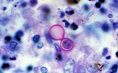
Blastomyces dermatitis is a biphasic fungus. The mycelial phase is formed at 22-30 °C, the mycelium is branching, septate, transverse, about 3 μm in size. Microconidia are round, oval or pear-shaped, 2x10 μm in size, attached to the lateral conidiophores. Lumpy chlamydospores are found in large quantities, resembling macroconidia of H. capsulatum and H. duboisii. At 37 °C and in an affected organism, the fungus is represented by a yeast phase. Yeast cells are large (10-20 μm), multinucleate, bear single buds attached to the mother cell by a wide base.
Cultural properties of Blastomyces dermatitis
Unpretentious to the nutrient substrate. At 25 °C it grows with the formation of hyaline (non-pigmented) hyphae with partitions and round or pear-shaped conidia, and at 37 °C it forms large thick-walled yeast cells with buds that are connected to the mother cell by a wide base.
Biochemical activity is low.
 [ 1 ], [ 2 ], [ 3 ], [ 4 ], [ 5 ], [ 6 ], [ 7 ]
Antigenic structure of Blastomyces dermatitis
When grown in liquid medium for 3 days, the mycediform produces exoantigen A, which can be determined using gel immunodiffusion and ELISA. Antigens A and B have been described for the yeast phase.
[ 8 ], [ 9 ], [ 10 ], [ 11 ], [ 12 ], [ 13 ]
Pathogenicity factors
Microconidia.
Ecological niche of Blastomyces dermatitis
Soil of endemic zones covering the USA (southern and south-central states), Canada (Great Lakes region), South America and Africa.
[ 14 ], [ 15 ], [ 16 ], [ 17 ], [ 18 ], [ 19 ]
Sustainability in the environment
It is not very stable in soil.
Antibiotic sensitivity
Sensitive to amphotericin B and ketoconazole.
Sensitivity to antiseptics and disinfectants
Sensitive to commonly used antiseptics and disinfectants.
Pathogenesis of blastomycosis
Microconidia enter the lungs, where primary foci of inflammation develop. Microconidia transform into large yeast cells. When granulomas form, areas of suppuration and necrosis are revealed, adjacent to intact tissues. Expressed alteration processes predetermine the massiveness of the fungus release with pathological material. There are cases of primary blastomycosis of the skin that developed after trauma. The development of mycosis is facilitated by diabetes mellitus, tuberculosis, hemoblasts, immunosuppressive states; in such individuals, blastomycosis shows a tendency to disseminaton. The disseminated (systemic) form of the disease can develop several years after the primary pulmonary lesion. Any organs can be removed in the pathological process, but the skin, bones, organs of the male genitourinary system, and adrenal glands are most often affected.
Cellular immunity
Its intensity and duration have not been studied.
Epidemiology of blastomycosis
The source of the infectious agent is the soil of endemic zones. Diseases of hunting dogs confirm the idea of the presence of the same sources of the pathogen for humans and animals. The mechanism of transmission is aerogenic, the route of transmission is airborne dust. Massive penetration of yeast cells leads to the introduction of the pathogen through the mucous membranes. Susceptibility of the population is universal, patients are not contagious to others. Low morbidity is explained by the small size of the fungal vegetation areas, which minimizes the risk of infection.
Symptoms of blastomycosis
The incubation period ranges from several weeks to 4 months. The disease may begin as a respiratory infection with minimal symptoms or acutely and be accompanied by a sudden increase in temperature, cough with purulent sputum, myalgia and arthralgia. Pneumonia often ends within 6-8 weeks without treatment. Subsequently, a number of such patients develop mycosis. Widespread pneumonia often leads to the death of the patient, despite timely treatment.
In the cutaneous form of the disease, the primary lesions are nodules, from which ulcers form. Ulceration areas with purulent discharge alternate with scarring zones. Ulcerative lesions can cover the mucous membrane of the oral cavity, spreading to the pharynx and larynx.
Laboratory diagnostics of blastomycosis
The materials examined include pus from fistulas and abscesses, cerebrospinal fluid, sputum, urine, and lymph node puncture.
Most often, a microscopic examination of pathological material is used. In a native preparation, clarified, round or oval large yeast cells with a double-contour cell wall are found, which form a single lobe with a wide base.
To isolate a pure culture, the material to be studied is sown on Sabouraud medium, sugar agar, or beer wort. The sown areas are incubated at 37°C to obtain yeast cells and at 25-30°C to obtain the initial phase. Transformation of yeast cells into mycelium is achieved by lowering the growth temperature to 25-30°C. Characteristic morphological elements of the mycelial phase are observed after 2-3 weeks of incubation. Smears from the fungal culture contain a capsule, a wide septate mycelium with thick walls. Conidia are round, oval, or pear-shaped. Chlamydospores are formed in old cultures.
The bioassay is carried out on white mice, followed by seeding of the affected tissue into nutrient media.
For serological diagnostics, RSC, ELISA, and RIA are used. Complement-fixing antibodies in sufficient titers are detected in the late stages of the disease.
Intradermal allergy tests are performed with the allergen blastomycin.