Medical expert of the article

New publications
Bronchiectatic disease - Causes and pathogenesis
Last reviewed: 04.07.2025

All iLive content is medically reviewed or fact checked to ensure as much factual accuracy as possible.
We have strict sourcing guidelines and only link to reputable media sites, academic research institutions and, whenever possible, medically peer reviewed studies. Note that the numbers in parentheses ([1], [2], etc.) are clickable links to these studies.
If you feel that any of our content is inaccurate, out-of-date, or otherwise questionable, please select it and press Ctrl + Enter.
Predisposing factors for the development of congenital bronchiectasis include smoking and alcohol consumption by the expectant mother during pregnancy and viral infections suffered during this period.
The development of bronchiectasis is facilitated by chronic diseases of the upper respiratory tract (sinusitis, chronic purulent tonsillitis, adenoids, etc.), which are observed in almost half of patients, especially in children.
Causes of Bronchiectasis
The causes of bronchiectasis have not yet been fully established. The most important etiological factors, proven to some extent, are the following.
- Genetically determined inferiority of the bronchial tree (congenital “weakness of the bronchial wall”, insufficient development of the smooth muscles of the bronchi, elastic and cartilaginous tissue, insufficiency of the bronchopulmonary defense system - see “ Chronic bronchitis ”), which leads to a disruption of the mechanical properties of the bronchial walls when they become infected.
- Infectious and inflammatory diseases of the bronchopulmonary system suffered in early childhood (often in older age groups), especially frequently recurring. They can be caused by various infectious agents, but the most important are staphylococci and streptococci, Haemophilus influenzae, anaerobic infection, etc. Of course, infectious and inflammatory diseases of the bronchopulmonary system cause the development of bronchiectasis in the presence of a genetically determined inferiority of the bronchial tree. Infectious agents also play a huge role in the development of exacerbations of the suppurative process in already altered and dilated bronchi.
- Congenital disorder of the development of the bronchi and their branching, which leads to the formation of congenital bronchiectasis. They are observed in only 6% of patients. Congenital bronchiectasis is also characteristic of Kartegener's syndrome (reverse arrangement of organs, bronchiectasis, sinusitis, immobility of the cilia of the ciliated epithelium, infertility in men due to a sharp impairment of sperm motility).
Bronchiectasis easily occurs in patients with congenital immunodeficiencies and congenital anatomical defects of the tracheobronchial tree (tracheobronchomegaly, tracheoesophageal fistula, etc.), with pulmonary artery aneurysm.
Bronchiectasis may accompany cystic fibrosis, a systemic, genetically determined disease with damage to the exocrine glands of the bronchopulmonary system and the gastrointestinal tract.
 [ 1 ], [ 2 ], [ 3 ], [ 4 ], [ 5 ], [ 6 ]
Pathogenesis of bronchiectasis
Pathogenesis includes factors leading to the development of bronchiectasis and factors leading to its infection. The development of bronchiectasis is caused by:
- obstructive atelectasis, which occurs when the bronchial patency is impaired (the development of atelectasis is facilitated by a decrease in surfactant activity, compression of the bronchi by hyperplastic hilar lymph nodes in the case of hilar pneumonia, tuberculous bronchoadenitis; long-term blockage of the bronchi by a dense mucous plug in acute respiratory infections). Obstruction of the bronchus causes a delay in the removal of bronchial secretions distal to the site of impaired bronchial patency and, of course, contributes to the development of irreversible changes in the mucous, submucous and deeper layers of the bronchial wall;
- decreased resistance of the bronchial walls to the action of bronchodilating forces (increased intrabronchial pressure during coughing, stretching of the bronchi by accumulating secretions, increased negative intrapleural pressure due to a decrease in the volume of the atelectatic part of the lung);
- the development of the inflammatory process in the bronchi, if it progresses, leads to degeneration of the cartilaginous plates, smooth muscle tissue with replacement by fibrous tissue and a decrease in the stability of the bronchi.
The following mechanisms lead to infection of bronchiectasis:
- impaired coughing, stagnation and infection of secretions in dilated bronchi;
- dysfunction of the local bronchopulmonary defense and immunity system.
According to A. I. Borohova and R. M. Paleev (1990), Klebsiella, Pseudomonas aeruginosa, Staphylococcus aureus, and less frequently Proteus and Streptococcus are most frequently found in the purulent contents of bronchiectasis. N. A. Mukhin (1993) points to the frequent detection of mycoplasma. In turn, the suppurative process in the bronchi promotes the expansion of the bronchi. Subsequently, the blood flow through the pulmonary arteries decreases, and the network of bronchial arteries hypertrophies, and blood is discharged from the bronchial arteries into the pulmonary artery system through extensive anastomoses, which leads to the development of pulmonary hypertension.
Pathomorphology
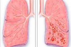
The bronchi of medium caliber are predominantly subject to expansion, less often - distal bronchi and bronchioles. Cylindrical, fusiform, saccular, mixed bronchiectasis is distinguished.
In cylindrical bronchiectasis, the expansion of the bronchi is moderately expressed, there is no significant deformation of the bronchial tree. Fusiform bronchiectasis is characterized by moderate expansion and deformation of the bronchi and a decrease in the number of bronchial deletions. Saccular bronchiectasis is the most severe form of bronchiectasis, with the proximal (central) bronchi initially affected, and as the disease progresses, expansion and then damage with subsequent fibrosis of the distal bronchi occur. As a result of these pathological processes, bronchiectasis in the peripheral sections is formed in the form of "bags" filled with pus.
Bronchiectasis is most often localized in the posterior basal segments of the lower lobes of both lungs and the middle lobe of the right lung.
The most characteristic pathomorphological manifestations of bronchiectasis are:
- dilation of the bronchi into a cylindrical or saccular shape;
- picture of a chronic purulent inflammatory process in the wall of dilated bronchi with pronounced peribronchial sclerosis;
- atrophy and metaplasia of the bronchial ciliated epithelium into multi-row or stratified squamous epithelium, in some places - replacement of the epithelium with granulation tissue;
- restructuring of the vascular network of the bronchi and lungs (opening of reserve capillaries; formation of arteriovenous anastomoses; hypertrophy of the muscular layer of the bronchial arteries and their expansion; formation of myoelastosis, myoelastofibrosis, elastofibrosis in the walls of the veins). The above changes in the arteries can be the cause of hemoptysis in bronchiectasis;
- changes in lung tissue in the form of atelectasis, pneumofibrosis and emphysema.