Medical expert of the article

New publications
Blood bivalve
Last reviewed: 04.07.2025

All iLive content is medically reviewed or fact checked to ensure as much factual accuracy as possible.
We have strict sourcing guidelines and only link to reputable media sites, academic research institutions and, whenever possible, medically peer reviewed studies. Note that the numbers in parentheses ([1], [2], etc.) are clickable links to these studies.
If you feel that any of our content is inaccurate, out-of-date, or otherwise questionable, please select it and press Ctrl + Enter.
The blood fluke or blood schistosome (Schistosoma haematobium) belongs to the parasites of the flatworm type (Phylum Plathelminthes), the class of flukes or trematodes (Trematoda Digenea), the order Strigeidida, the family Schistosomatidae.
S. haematobium infections remain a major public health problem in most countries of Africa and the Middle East, second only to malaria among parasitic diseases.
 [
[ Epidemiology
According to WHO statistics, 180 million people worldwide live in endemic areas and 90 million are infected with the parasite. It is estimated that almost 150,000 people die each year due to complications of urogenital schistosomiasis; the overall mortality rate is 2 per 1,000 infected patients per year.
Causes blood bivalve
It should be noted that the blood fluke is a bisexual worm, coexisting in a male-female pair. Accordingly, their structure is somewhat different. The length of the wider tubular body of the male does not exceed 10-15 mm, while the narrower body of the female can be 2 cm long. Each male has a unique gynecophoral canal on the abdominal part, in which his female is constantly located.
There are suckers on the front and abdominal parts of the body, the female has an ovary with an oviduct leading to the genital opening behind the intestinal canal. The size of the oval eggs in length is about 0.15 mm, on one side the eggs have a pointed shape with a thorn. Inside the eggs are larvae - miracidia.
Pathogenesis
The blood fluke is infectious to humans and causes the parasitic disease urogenital schistosomiasis, which can lead to the development of pathological neoplasms.
Structure and life cycle of the blood fluke
The life cycle of the blood fluke occurs in the organisms of two hosts. The intermediate host is freshwater gastropods (snails) of the Planorbidae family, genus Bulinuss, living in the waters of Africa and the Middle East. The final host is man.
The first larval stage begins when miracidia of 0.2 mm in size emerge from the eggs that fall into the water. They have excretory organs (two pairs of protonephridia) and cilia on the outside that allow free movement in the water. When the miracidia enter the snail's body, they asexually divide intensively and form two generations of sporocyst larvae. The sporocyst structure is normal, in the form of a pleomorphic body (sac) containing developing larvae. Cercariae, the third larval stage of the blood fluke, develop from the daughter sporocysts within 2-3 weeks. Growing to approximately 0.3 mm, the cercariae leave the snail's body and end up in the water again. This is an invasive form, since the cercaria has a forked tail (furcocercous) and moves quickly in search of a definitive host.
The routes of human infection are the introduction of cercariae through the skin into the body (when a person comes into contact with stagnant or slowly flowing water) and their penetration into the blood. Parasitologists do not rule out infection when water enters the gastrointestinal tract through the mouth.
The cercariae drop their tail and transform into schistosomes, which enter the mesenteric venules of the abdominal cavity, rectal venules, and venous plexus of the urinary bladder with the blood flow. Here, each schistosome undergoes a sequential transformation into an adult paired worm, which attaches to the wall of the vessel with its abdominal sucker and feeds on blood through its oral sucker.
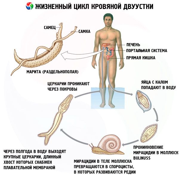
After 4-8 weeks of infection, female S. haematobium begin laying eggs (200-3000 per day), which move progressively towards the bladder and ureters and, perforating the wall, penetrate the bladder. During urination, the eggs come out and end up in the water. And a new life cycle of the blood fluke begins. Adult worms usually live for 2-5 years, although some can live much longer.
Symptoms blood bivalve
Not all eggs penetrate the bladder, many of them end up in the organs with the bloodstream, where they form characteristic granulomas in the form of polyps surrounded by inflammatory cells. After the death of the encapsulated eggs, the granulomas harden, causing various pathologies of the internal organs.
Urogenital schistosomiasis, which is caused by a blood fluke, does not develop immediately. Early symptoms of infection with this parasite appear approximately 24 hours after the fluke penetrates: an itchy papular rash and local swelling appear on the skin at this site. This period lasts about 4-5 days.
For one to two months, symptoms of infection may include fever, enlarged liver, spleen, and lymph nodes. During this period, which lasts an average of one to three weeks, anemia, an increase in the number of eosinophilic leukocytes in the blood (eosinophilia), or a decrease in the level of platelets are observed. However, as doctors note, not everyone shows signs at an early stage of the disease, and the course of the disease is also individual.
After several months or even years, 50-70% of those infected may experience pain when urinating and dysuria, blood appears in the urine (hematuria); urethral obstruction and kidney damage in the form of obstructive nephropathy also develop.
With dysfunction of the urinary tract caused by the blood fluke, hydronephrosis (accumulation of urine in the kidneys) develops; any bacterial infection may also join in, which leads to the development of cystitis - with the corresponding symptoms. During an endoscopic examination of the bladder, granulomas (clusters of S. haematobium eggs), polyps, ulcers, areas of calcification or keratinization of the mucous membrane (leukoplakia) are detected. During the examination of women with schistosome invasion, focal growths of the vaginal or cervical mucosa, urethral fistulas are detected. Intestinal polyposis, pulmonary arteritis, cardiovascular problems, including heart failure and periportal fibrosis may also develop.
Diagnostics blood bivalve
Diagnosis of blood fluke includes anamnesis (the patient must report visiting endemic areas) and urine analysis (eggs of the parasite are detected in it). The eggs are a fairly characteristic diagnostic sign. In some cases, a biopsy of the bladder, rectum or vaginal wall may be used.
Who to contact?
Treatment blood bivalve
Typically, treatment for blood fluke is carried out using drugs such as:
- Biltricide (Praziquantel): single oral dose is calculated based on body weight. 20 mg/kg three times during one day or a single dose of 40 mg per kilogram of weight.
- Metrifonate: taken for three weeks - once a week at 10 mg per kilogram of body weight.
- Hicanton (Etrenol): administered intramuscularly once, the dose is determined based on 2-3 mg per kilogram of weight.
Corrective surgery may be necessary in cases of urinary tract obstruction. Complications of urogenital schistosomiasis should be treated with appropriate methods and drugs.
Prevention
Prevention of infection with the blood fluke and the development of urogenital schistosomiasis is a pressing problem for endemic regions, which include more than 50 countries in Africa and the Middle East.
The blood fluke parasitizes mainly among the inhabitants of rural areas of these regions, where snails (intermediate hosts of the trematode) also live; many human activities also affect the distribution of the parasite, especially the construction of irrigation canals and irrigation systems.
Preventive measures include improved sanitation, biological control of the snail population that carries the blood fluke, and the use of molluscicides to combat them. Education of local populations and tourists visiting endemic areas plays an important role.