Medical expert of the article

New publications
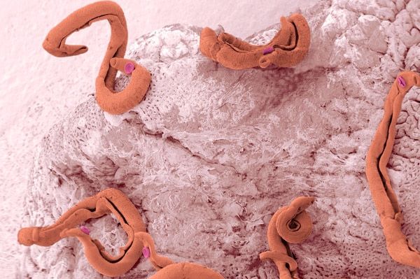
Schistosomes
Last reviewed: 06.07.2025

All iLive content is medically reviewed or fact checked to ensure as much factual accuracy as possible.
We have strict sourcing guidelines and only link to reputable media sites, academic research institutions and, whenever possible, medically peer reviewed studies. Note that the numbers in parentheses ([1], [2], etc.) are clickable links to these studies.
If you feel that any of our content is inaccurate, out-of-date, or otherwise questionable, please select it and press Ctrl + Enter.
Schistosomes are parasites from the group of flatworms or trematodes, which are also called blood flukes. They are one of the most harmful flukes due to possible complications, so it is very important to identify the pathogen in time and carry out treatment.
Features of the structure and life cycle of different types of schistosomes
Schistosomes belong to the class of trematodes, which characterizes them as individuals with a complex life cycle. They have several hosts and their life cycle takes place with the participation of freshwater mollusks. These are individuals of different sexes, but they have the ability to merge at some point and the male carries the female on his body. Therefore, the structure of the male is such that he is slightly shorter than the female in length, but he is thicker. The female has a long flat body. After merging, she is in a special sac of the male - the gynecoform canal. The pathogen is also called blood schistosome due to its predominant localization in the vessels of the human body.
There are several species of schistosomes that are pathogenic to humans.
Schistosoma mansoni is a parasite that causes intestinal schistosomiasis. It is characterized by damage to the veins or venules of the intestine, mainly the mesenteric vessels. This leads not only to mechanical damage to the intestinal wall, but also to a toxic effect on the functioning of the intestine. Therefore, a feature of this type of schistosome is the predominance of intestinal symptoms.
The urogenital schistosome or schistosome hematobium is the causative agent of urogenital schistosomiasis. This species has a body covered with spines, which allows it to attach to the mucous membrane and withstand the action of urine. This parasite is localized in the veins of the small pelvis - the veins of the uterus, bladder, and is also highly mutagenic. It causes symptoms that are characteristic of the localization of the pathogen - urination and sexual dysfunction, as well as menstrual dysfunction.
Japanese schistosome is also the causative agent of the intestinal form, but it has a more severe course and is common in the regions of Indonesia, Japan and China. The peculiarity of the pathology is the rapid course and progression of the disease, which can cause a rapid deterioration in the condition and progressive liver dysfunction up to cirrhosis.
The size of the male schistosome is about ten to fifteen centimeters, and the female is more than twenty centimeters. They live separately for several months, then they unite and the male carries the female for the rest of her life. Males have a sucker with which they can attach themselves to the inner wall of a vessel and actively move.
The parasite's life cycle begins with the eggs produced by a mature female being excreted into the environment with feces or urine. For further development, they must enter fresh water, where their intermediate host is located. The eggs are swallowed by mollusks of different genera, where further development and formation of larvae occurs. Schistosoma larvae emerge from the mollusk and are able to actively swim in the water.
The routes of infection with schistosomes are contact. They penetrate the human body when swimming in a pond, even through clothes, as well as with accidental swallowing of water or intentional consumption of water. Then, after entering the human body, the larva penetrates the vessels and actively migrates throughout the body. The location of the final localization of the parasite depends on the type of pathogen and its tropism is determined by tropism to certain organs. Then, after activation in the veins of certain organs, the parasite can live up to forty years, while a toxin is released and the corresponding symptoms appear. There are also local manifestations due to damage to the organ wall and disruption of its trophism and venous outflow.
Symptoms of Schistosomiasis Infection
Clinical symptoms of schistosomiasis infection can be divided into several stages - pre-hepatic, hepatic and post-hepatic. The incubation period is from three to six weeks. When the larva enters the human body, it enters the liver from the intestines or from the skin vessels at the larval stage. This is the pre-hepatic stage of the parasite's development. If the schistosome has penetrated the skin, a pinpoint rash, itching, and burning in the area appear at the site of penetration.
Further, at the penetration stage, there may be an allergic reaction throughout the body in the form of a polymorphic rash on the skin, like urticaria. The acute period of the disease, which corresponds to the migration of the parasite through the venous vessels of the body, is characterized by non-specific manifestations in the form of increased body temperature, pain in muscles and joints, and sleep disorders.
In the liver stage, the parasite grows and reproduces in the portal vein system, which corresponds to the next stage of development. The posthepatic stage of development is characterized by further migration of the male with the female and localization in the pelvic vein system. This corresponds to the stage of widespread invasion, in which the sexually mature parasite actively moves directly in the organs and lays eggs.
Further, two weeks after the onset of the disease, specific symptoms from the intestines or genitourinary system may appear.
Symptoms from the genitourinary system occur due to the pathogenic action of schistosome. Mechanical action occurs when the eggs damage the walls of the genitourinary organs - in this case, erosions, ulcers, signs of inflammation and polypous reactions are observed on the mucous membrane of the bladder due to long-term parasitism. There is also a toxic-allergic effect due to the constant vital activity of schistosome and the release of metabolic products into human blood. Trophic processes of the bladder and uterus are disrupted, which causes a violation of cell division and is a risk factor for the development of oncopathology. The parasite also feeds on erythrocytes and nutrients, which significantly disrupts the general trophism of the human body and the respiratory function of the blood.
There may also be general symptoms when the parasite migrates and localizes in the lungs - a paroxysmal cough, shortness of breath, difficulty breathing. These symptoms disappear when the parasite moves to its final location. Local symptoms mainly manifest as urination problems, pain during urination, and hematuria (the appearance of blood in the urine). If the schistosome is localized in the veins of the uterus or in the extrauterine space, then there may be pain in the lower abdomen not associated with menstruation, menstrual cycle disorders.
In chronic cases, complications are often observed - ureteral strictures, pyelonephritis, hydronephrosis, as well as the formation of stones in the kidneys and bladder. Schistosomes can cause early impotence.
Schistosoma Mansoni, when entering the body in the acute stage, also has similar symptoms of general intoxication, muscle pain and skin rash. Further, given its localization, symptoms of dyspepsia will be expressed. At first, clinical manifestations in the form of abdominal pain, stool disorders such as diarrhea. Then, with an increase in the number of parasites and eggs, strong mechanical irritation occurs and this leads to the fact that diarrhea alternates with constipation, there may be mucus and blood in the feces. Tenesmus occurs and this can lead to bleeding and even prolapse of the rectum.
Japanese schistosome is characterized by intestinal symptoms that have a strong pronounced course with predominant damage to the liver. In this case, the structure of hepatocytes is disrupted, which leads to the rapid development of liver cirrhosis. Therefore, along with intestinal manifestations, symptoms are also observed in the form of an enlarged liver, its soreness upon palpation, yellow color of the sclera and skin.
Diagnosis of Schistosoma
Schistosomiasis diagnostics is much easier if there is epidemiological data on swimming in a body of water or contact with a source of infection. Anamnesis data allows identifying the first symptoms of the pathology and studying the course of the disease.
Analysis for schistosomes is carried out taking into account the localization of the pathological process. If the patient complains of the genitourinary system, it is necessary to conduct a microscopic analysis of urine - this reveals schistosome eggs. During ovoscopy, you can see schistosome eggs, which are oval in shape, elongated, with spikes on one side. With instrumental methods, it is sometimes necessary to conduct a cystoscopy. In this case, you can see erosions on the inner mucous membrane of the bladder, signs of inflammation. In biopsy samples, you can determine the pathogen itself, its eggs, as well as signs of damage to the integrity of the wall.
In intestinal schistosomiasis, the diagnosis can be confirmed by examining feces and microscopy of feces. If there are signs of mucus and blood in the feces, then a rectoscopy is performed, which allows examining the intestinal mucosa and taking a biopsy. In biopsies, it is possible to identify the pathogen or its eggs, which makes it possible to exclude autoimmune intestinal damage (nonspecific ulcerative colitis). A general blood test is mandatory. Specific changes that may indicate helminthic invasion include blood eosinophilia. This also indicates activation of the allergic link of the immune system. In the biochemical blood test, there may also be changes in the acute stage in the form of an increase in liver enzymes (alkaline phosphatase), as well as hyperbilirubinemia of mixed genesis, especially if we are talking about Japanese schistosome, which affects the liver.
Immunological methods are also used for diagnostic purposes. To do this, the presence of antibodies in the patient's body is determined using the indirect hemagglutination reaction. The most reliable method is considered to be the determination of the genetic material of the schistosome in the patient's feces, blood, urine or other biological fluid. For this, a polymerase chain reaction is used, which allows for the accurate determination of the parasite's DNA and confirmation of the diagnosis.
 [ 11 ], [ 12 ], [ 13 ], [ 14 ], [ 15 ], [ 16 ], [ 17 ], [ 18 ], [ 19 ], [ 20 ], [ 21 ]
Treatment of schistosomiasis
Treatment of schistosomes should be carried out in the acute period, when the parasite is in the portal vein system and has not yet reached its target and is not localized in the pelvic organs or intestines. In this case, the use of specific antihelminthic drugs is most justified.
- Ambilgar is an antiparasitic agent, the active substance of which is niridazole. The drug has an active effect on schistosomes, both at the stage of invasion and at organ localization. The drug is available in the form of tablets of 100 milligrams and 500 milligrams and is dosed at 25 milligrams per kilogram of the patient's body weight. A side effect during the administration of the drug is possible in the form of increased excitability, drowsiness, and it is also possible to affect the hematopoietic system with the suppression of all germs.
- Baltricid is an antihelminthic drug, the active substance of which is praziquantel. The drug is highly effective against trematodes, including schistosomes. The mechanism of action is the activation of cellular channels by the drug, which increase the concentration of calcium inside - this leads to the fact that there is a strong contraction of the parasite's body without relaxation, and it dies. This drug is available in the form of tablets of 600 milligrams, the dosage of the drug is 25 milligrams per kilogram of the patient's body weight per day. Side effects are possible during the administration of the drug with a strong helminthic invasion - nausea, abdominal pain, itching of the skin, as well as pronounced intoxication symptoms.
Symptomatic treatment is also necessary. In case of severe symptoms of intestinal toxoplasmosis, it is necessary to prescribe antispasmodics (Baralgin, Drotaverine), probiotics (Yogurt, Enterol, Lactiale), and an antidiarrheal agent. It is important to follow a diet to replenish energy deficiency and prevent intestinal symptoms.
Prevention
Prevention of schistosomiasis should be carried out in areas with an epidemiological situation for this disease. It is necessary to inform people about this disease, about the ways of its transmission, and to carry out sanitary treatment in the sources of active infection. Patients should be treated and possible ways of transmission of infection should be sanitized. If there is data on contact with a body of water and similar clinical manifestations, then it is necessary to carry out specific prevention using anthelmintic agents for therapeutic or preventive purposes.
Schistosomes are parasites that infect humans through contact with contaminated water when they enter through the skin or intestines. They have a very wide migration path through the body, which can cause many pathological symptoms. At the same time, schistosomes feed on red blood cells, and when localized in the genitourinary system or intestines, they lead to mechanical and toxic effects. Treatment of schistosomiasis is problematic, and given the serious complications, it is necessary to prevent this pathology.