Medical expert of the article
New publications
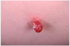
Benign granuloma on a pedicle.
Last reviewed: 04.07.2025

All iLive content is medically reviewed or fact checked to ensure as much factual accuracy as possible.
We have strict sourcing guidelines and only link to reputable media sites, academic research institutions and, whenever possible, medically peer reviewed studies. Note that the numbers in parentheses ([1], [2], etc.) are clickable links to these studies.
If you feel that any of our content is inaccurate, out-of-date, or otherwise questionable, please select it and press Ctrl + Enter.

Causes benign granuloma
Some scientists believe that benign granuloma is a specific form of pyoderma. Some dermatologists consider it a capillary hemangioma with a secondary granulomatous reaction. In recent years, it has been suggested that the disease is based on angioblastoma, which is joined by a bacterial infection.
Pathogenesis
In the pathogenesis of benign granuloma, trauma plays a major role - a cut, injection, burn, etc.
Histopathology. In the early stages of the disease, no signs of inflammation are observed in the epidermis, while in the later stages there are signs of destruction. In the dermis, a focus of a large number of newly formed vessels with swollen endothelium is observed. The infiltrate consists of polymorphic leukocytes, plasma cells, lymphocytes, and mast cells.
Symptoms benign granuloma
After a few weeks, a painless vascular tumor the size of a pea or cherry appears at the site of the injury, often as if sitting on a narrow or wide stalk, surrounded by a "collar" of exfoliated epidermis. The tumor is dark red in color with a smooth or lobular surface and has a dense, elastic consistency. Later, the elements bleed easily, partially ulcerate and become covered with bloody-purulent discharge.
The tumor is most often located on the hands, feet, face, but can also be localized in other areas of the skin. Regional lymph nodes, as a rule, are not involved in the process, except for rare cases of secondary infection. Sometimes the tumor-like formation has a wide infiltrated base of a round or oval shape. Multiple benign granulomas are rare.
 [ 11 ]
What do need to examine?
How to examine?
Differential diagnosis
Differential diagnosis is carried out with keratoacanthoma, cavernous angioma, Kaposi's sarcoma, angiosarcoma, molluscum contagiosum, pyoderma vegetans, seborrheic keratosis.
Who to contact?
Treatment benign granuloma
Surgical excision, electrocoagulation, and laser irradiation are performed.

