Medical expert of the article

New publications
Balantidia
Last reviewed: 06.07.2025

All iLive content is medically reviewed or fact checked to ensure as much factual accuracy as possible.
We have strict sourcing guidelines and only link to reputable media sites, academic research institutions and, whenever possible, medically peer reviewed studies. Note that the numbers in parentheses ([1], [2], etc.) are clickable links to these studies.
If you feel that any of our content is inaccurate, out-of-date, or otherwise questionable, please select it and press Ctrl + Enter.
Among the parasites living in humans, there are many different species. One of them is Balantidia, which lives in the large intestine of the human body. They belong to the group of infusoria and are also found in the bodies of some mammals. Pigs, rats, and dogs are the same carriers of Balantidia as humans.
This type of protozoa causes a disease in the body of its "host" called balantidiasis or infusoria dysentery. At the same time, balantidia are currently the only type of parasitic infusoria that have been proven to live in humans. The discoverer of the species was the Swedish scientist Malmsten, who described balantidia in 1857. But the parasitic effect of these infusoria on humans was discovered by the scientist N.S. Solovyov in 1901.
Despite the progress of modern medicine, balantidiasis is still detected in quite rare cases. Although carriers of the disease are considered to be about four to five percent of rural residents. Most often, among such potential patients there are people who, by their occupation, look after pigs. Pigs are natural carriers of balantidia and can infect humans with them through close contact. Infection of humans from humans occurs through contact between a healthy person and a sick person.
Currently, the following specialists are involved in the detection and treatment of diseases caused by balantidia: parasitologists and infectious disease specialists. It is very important not only to diagnose parasitic infection in time, but also to begin its therapy at the appropriate time. No less important are measures to prevent balantidia infections, which consist of the constant use of hygienic measures when interacting with pigs. In order to prevent the mass spread of parasitic diseases, it is necessary to carry out campaigns to identify and treat carriers of balantidia and patients with balantiasis.
 [
[ Structure of balantidia
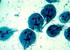
Balantidia belong to the protozoan type and the ciliate class due to the cilia that cover the entire body of the ciliate. In addition, this type of ciliate is considered the largest type of protozoan that lives in the human large intestine.
The structure of balantidia is as follows: the vegetative form of the cell is distinguished by its size from fifty to eighty microns by thirty-five to sixty microns. At the same time, the body of the ciliate is elongated, most often resembling an egg. In length, balantidia reaches from thirty to one hundred and fifty microns, and in width - from twenty to one hundred and ten microns.
The shape of this type of ciliates is ovoid, and the surface of the protozoa is covered with a pellicle. The pellicle has many short cilia, which are located longitudinally in a large number of rows. These cilia are organelles of movement that help the ciliate move. At the same time, the ciliate can not only actively move, but also describe rotations around its axis.
The pellicle of Balantidia is elastic, especially when it moves, so the symmetry of the ciliate's body can be disrupted when moving. Under the pellicle there is a thin layer of transparent alveolar ectoplasm.
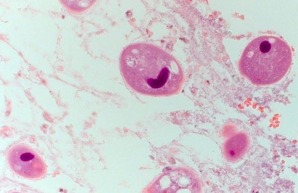
At the front end of the ciliate, a slit-like depression can be found, which is called the peristome. At the bottom of the depression, there is a mouth opening, called the cyostome. The structure of the nuclear apparatus of balantidia is no different from other ciliates and is represented by a macronucleus and a micronucleus. The nucleus - the so-called macronucleus - can be seen through the body membranes of some living individuals. It resembles a light bubble, which has a bean-shaped form.
In the cytoplasm of the protozoan there are digestive and pulsating vacuoles in the amount of two pieces. The pulsating vacuoles are excretory and through them the products of the vital activity of the ciliate are extracted.
The parasite feeds by absorbing food particles, such as starch grains. Blood cells are also suitable - erythrocytes and so on. In addition, balantidia feed on various bacteria and fungi. Nutrients are absorbed in contractile (pulsating) and digestive vacuoles, where all elements suitable for this purpose fall.
The cyst is round in shape and is from fifty to seventy microns in diameter. It is covered with a thick membrane. The cytoplasm inside the cyst is homogeneous.
Infusoria balantidia
Among the parasitic protozoa that live in humans, the ciliate balantidia is considered the largest species. Most often, this type of ciliate affects people living in the southern regions of the globe, especially where pig farming is widespread. Although, it is known that in sporadic forms, these protozoa have been identified everywhere where pig farming takes place.
It is very important to follow preventive measures to avoid contracting balantidiasis caused by balantidia. Personal hygiene rules should be relevant, especially when working with pigs. At the same time, it is equally important to use clean water for drinking, cooking and washing dishes, which has been purified using modern methods. It is also important to eat only clean and well-washed food products, vegetables and fruits, stored in compliance with all sanitary conditions.
Balantidium intestinalis
This type of protozoa lives exclusively in the human intestine. There it also causes various lesions of the mucous membrane of the large intestine. Therefore, in some cases this type of infusoria is called "intestinal balantidia". This name is common and is used by people who are not related to medicine.
Balantidia intestinalis is the same ciliates that were described earlier, only named differently. Therefore, to get acquainted with the structure of the simplest, as well as the features of its vital activity, we recommend referring to the previous sections of the article.
Life cycle of Balantidia
Like any other type of ciliates, balantidia have a certain cyclicity in their existence. The life cycle of balantidia consists of sexual and asexual phases. The sexual phase is also divided into sexual reproduction: conjugation, characterized by the exchange of nuclei between two representatives of balantidia and asexual reproduction, which is expressed in the transverse division of ciliates.
When the period of sexual reproduction ends, this type of protozoa turns into a cyst and in this form, most often, leaves the human body and is released into the environment along with feces. The cyst has no cilia, and it itself is covered with a membrane consisting of two layers. Such cysts can be viable for a long time, even without being in a living organism. In feces, if the temperature is room temperature, cysts can survive for up to thirty hours. Being in tap and waste water increases the viability of cysts to a week.
If balantidia cysts get on any objects from the environment, they can survive on them for up to two months. The main condition for their viability is that the temperature of the atmosphere should be close to room temperature, and the humidity should be increased. In dry and dark places, cysts survive for up to two weeks.
In some solutions it is possible to preserve balantidium cysts, but only for a short period of time. For example, a five percent aqueous solution of carbolic acid can help to prolong the viability of cysts for only three hours, and a formalin solution for four hours. It is possible to cultivate balantidium cysts in various nutrient media, which are organized for them in laboratory conditions.
Diseases that cause balantidia
The main disease that a person begins to suffer from due to infection with balantidia is called balantidiasis. This is a type of parasitic disease characterized by lesions of the mucous membrane of the large intestine, which appear in the form of ulcers. Sometimes balantidia spend their life activity not only in the large intestine, but also in the distal part of the small intestine. At first, ciliates penetrate the intestinal epithelium, and then begin to actively multiply in it. Such activity of protozoa causes an inflammatory-ulcerative process in the intestine. In this case, the disease is characterized by severe symptoms and high mortality of patients if treatment is not provided in a timely manner.
Symptoms of balantidiasis include:
- presence of diarrhea,
- the appearance of pain in the abdominal area,
- the occurrence of general intoxication of the body,
- the appearance of vomiting,
- the occurrence of headaches,
- the presence of mucus and blood in the patient's feces.
By the nature of the course, balantidiasis is divided into subclinical or latent (carrying cysts), acute and chronic, having a recurring nature. The chronic form of the disease is most often detected. Of the forms of the disease, moderate and severe are most often encountered. Also, such types of the disease are found that are combined with other parasitic infections - with amoebiasis, and shigellosis, and so on.
The incubation period for balantidiasis is ten to fifteen days. However, there are known cases where the incubation period was five to thirty days.
Both acute and chronic balantidiasis proceed as follows. The patient may develop one of the forms of the disease:
- balantid dysentery, which causes foul-smelling, bloody-red diarrhea,
- balantid colitis, which manifests itself in the form of semi-liquid feces with mucus impurities, but without blood inclusions.
Balantid dysentery in its acute form, in which the patient was not given timely specific treatment, leads to frequent cases of death.
If balantidiasis is not complicated by another bacterial infection, then in this case, especially in the acute stages of the disease, the patient does not have an elevated body temperature. Also, the disease is not characterized by complications that affect other organs of the human body.
The course of the disease in its acute form is as follows. The symptoms of the disease resemble those of enterocolitis or colitis. In this case, patients begin to feel general intoxication of the body: weakness and headache, loss of appetite. In half of the cases, acute balantidiasis is accompanied by moderate fever and sometimes chills. At the same time, symptoms of intestinal damage are present: abdominal pain, diarrhea, flatulence. If the rectum is also involved in the inflammatory and ulcerative process, tenesmus may appear - false urges to defecate. Mucous and blood impurities are characteristic of feces. Sometimes patients experience dryness and coating of the tongue, as well as spasms and painful sensations in the large intestine itself. The liver becomes painful and enlarged.
Rectomanoscopy examination always reveals the presence of a focal infiltrative-ulcerative process. Laboratory blood tests reveal signs of moderate anemia, eosinophilia, and decreased levels of proteins and albumins. ESR becomes moderately increased.
If acute balantidiasis has a severe course, the patient experiences the following symptoms: high fever, sharp symptoms of intoxication, in which the patient experiences signs of chills, nausea, vomiting and headache. Bowel movements can be up to twenty times a day, while they contain mucus and blood, and the smell of feces becomes putrid. Patients lose a lot of weight, and after a week, cachexia can be diagnosed. Sometimes signs of peritoneal irritation appear.
The rectoscopy procedure in this case reveals the presence of extensive ulcerative changes in the mucous epithelium of the large intestine. Laboratory blood tests reveal the presence of hypochromic anemia and neutrophilic leukocytosis.
The chronic form of the disease manifests itself as follows: exacerbation phases are characteristic, which are similar to acute balantidiasis, as well as periods of remission. At the same time, during remissions, symptoms of the disease may be absent, including diarrhea.
In chronic balantidiasis, signs of intoxication are expressed in a weak form, and the body temperature remains normal. Defecation occurs about two or three times a day, the stool becomes liquid, with admixtures of mucus, and sometimes - blood. Palpation reveals painful sensations in the area of the cecum and ascending colon.
Diagnostic procedures using rectoscopy confirm the presence of typical ulcerative changes in the intestinal mucosa. And the diagnosis is confirmed by laboratory tests of feces, which reveal parasites.
Diseases that cause balantidia are most often life-threatening, so it is very important to correctly diagnose balantidiasis and begin appropriate treatment in a timely manner.
Treatment of Balantidia
Before carrying out therapy for any disease, it is necessary to conduct a high-quality diagnosis, which will allow you to verify the presence of a certain disease.
Balantidiasis is diagnosed as follows. A drop of feces that has just been excreted must be placed in an isotonic solution of sodium chloride. All of the above is placed on a glass slide and examined using a microscope. Balantidia can be detected due to their large size and active movement.
The release of infusoria occurs periodically, so diagnostics should be carried out not once, but several times, to really make sure of the presence of these parasites. Sometimes, in order to conduct a study of the patient's feces, he is prescribed the use of a saline laxative. It is necessary to know that carriers of balantidia have only single cysts, which are difficult to detect.
Balantidiasis, as a parasitic disease, requires effective therapy. Treatment of balantidia is successfully carried out using etiotropic methods, which include the use of certain drugs, namely:
- Metronidazole or Trichopolum.
Adult patients should take 1.2 grams of the drug per day, and children - 0.75 grams of the drug. The course of treatment is seven days.
- Monomycin.
Adults take a dose of the drug, which is from fifty thousand to two hundred and fifty thousand units, four times a day. The course of treatment is five days with a break from five days to one week. Then the five-day course of therapy must be repeated.
In severe forms of the disease, the course of treatment consists of three five-day doses of the drug with two breaks of five to seven days.
- Tetracycline.
This medication is prescribed for severe manifestations of the disease. Adults take two grams of the drug per day for a week.
- Diyodokhin.
- Yatren.
Also, in parallel with the above therapy, it is necessary to carry out detoxification and non-specific stimulating treatment of the disease.
The patient's recovery is confirmed by specialists if the patient does not have colitis syndrome. Also important are the data of coprological examination and intestinal wall reparation, in which the absence of balantidia is observed.
Balantidia are serious violators of human well-being and health. Therefore, at the slightest strange symptoms indicating parasitic infestation, it is recommended to undergo a comprehensive examination to identify protozoa in the body. In this case, human health can be restored in the shortest possible time, which will save him from serious consequences and problems.