Medical expert of the article

New publications
Auditory (eustachian) tube
Last reviewed: 07.07.2025

All iLive content is medically reviewed or fact checked to ensure as much factual accuracy as possible.
We have strict sourcing guidelines and only link to reputable media sites, academic research institutions and, whenever possible, medically peer reviewed studies. Note that the numbers in parentheses ([1], [2], etc.) are clickable links to these studies.
If you feel that any of our content is inaccurate, out-of-date, or otherwise questionable, please select it and press Ctrl + Enter.
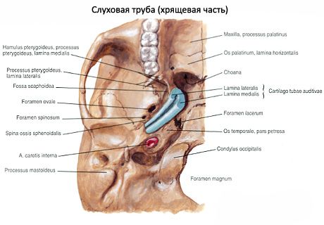
The auditory (Eustachian) tube (tuba auditiva, s. auditoria) is on average 35 mm long and 2 mm wide. Through it, air from the pharynx enters the tympanic cavity to maintain the pressure in the cavity equal to the external pressure, which is important for the normal functioning of the sound-conducting apparatus (eardrum and auditory ossicles). The auditory tube is divided into a bony part (pars ossea) and a cartilaginous part (pars cartilaginea), consisting of elastic cartilage. The lumen of the tube at the junction - the isthmus of the auditory tube (isthmus tubae auditivae), narrows to 1 mm. The upper bony part of the tube is located in the hemichannel of the same name of the muscular-tubular canal of the temporal bone and opens on the anterior wall of the tympanic cavity by the tympanic opening of the auditory tube (ostium tympanicum tubae auditivae). The lower cartilaginous part, which accounts for 2/3 of the length of the tube, has the form of a groove, open at the bottom, formed by the medial and lateral cartilaginous plates and the membranous plate connecting them. In the place where the auditory tube opens on the lateral wall of the nasopharynx with the pharyngeal opening of the auditory tube (ostium pharyngeum tubae auditivae), the medial (posterior) plate of the elastic cartilage of the tube thickens and protrudes into the cavity of the pharynx in the form of a tubular ridge (torus tubarius). The longitudinal axis of the auditory tube from its pharyngeal opening is directed upward and laterally, forming an angle of 40-45° with the horizontal and sagittal planes.
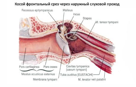
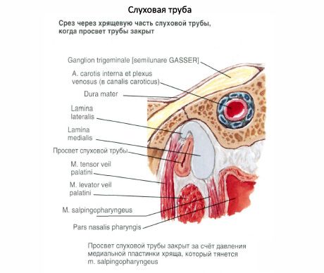
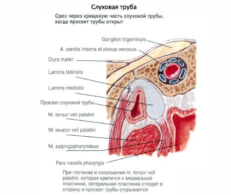
The muscle that tenses the soft palate and the muscle that lifts the soft palate originate on the cartilaginous part of the auditory tube. When they contract, the cartilage of the tube and its membranous plate (lamina membranacea) are pulled back, the canal of the tube expands and air from the pharynx enters the tympanic cavity. The mucous membrane of the tube forms longitudinal folds and is covered with ciliated epithelium, the movements of the cilia of which are directed towards the pharynx. In the mucous membrane of the auditory tube there are many mucous tubal glands (glandulae tubariae) and lymphoid tissue, which near the tubal ridge and around the pharyngeal opening of the auditory tube forms a cluster - the tubal tonsil (see "Organs of hematopoiesis and the immune system").
 [
[ Where does it hurt?
What do need to examine?
How to examine?