Medical expert of the article

New publications
Ear examination
Last reviewed: 06.07.2025

All iLive content is medically reviewed or fact checked to ensure as much factual accuracy as possible.
We have strict sourcing guidelines and only link to reputable media sites, academic research institutions and, whenever possible, medically peer reviewed studies. Note that the numbers in parentheses ([1], [2], etc.) are clickable links to these studies.
If you feel that any of our content is inaccurate, out-of-date, or otherwise questionable, please select it and press Ctrl + Enter.
Doctors working in the otolaryngology department are always very noticeable: above their eyes there is always a concave mirror with a hole in the center. These are reflectors that collect rays from an independent light source into a strong beam that perfectly illuminates the ENT organs, allowing them to be examined stereoscopically, while leaving the hands free for manipulation.
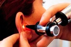
How is an ear examination performed?
First of all, examine the auricle and adjacent areas for inflammation and swelling. If there is discharge from the ear, take a swab for culture and remove wax from the external auditory canal. Attach the most convenient and largest ear funnel to the otoscope and examine the external auditory canal and eardrum as follows. Pull the auricle upward and backward, thereby straightening the external auditory canal (in infants, the auricle should be pulled downward and backward). The handle of the malleus is a good landmark located behind the eardrum. Anteriorly and posteriorly, you can see a good light reflex formed in this place due to the concavity of the eardrum. It is necessary to note the transparency of the eardrum, its color, and whether it is bulging or perforated. Perforation of the eardrum in its relaxed part indicates a serious pathology. The mobility of the eardrum can be tested using a funnel with a piece of glass covering the front and a small "tip" on the side to which a small rubber bulb is attached. As you squeeze the bulb, the eardrum begins to move. The Eustachian tube can be seen as the eardrum moves when the patient performs the Valsalva maneuver.
Anatomy of the ear
The cartilage of the auricle develops from six tubercles. If its sections do not tightly merge with each other during development, fistulas (most often a small fistula in front of the tragus) or accessory auricles (cartilaginous bodies located between the corner of the mouth and the tragus) may form.
The external auditory canal is 3-4 cm long and has a slightly S-shape. The outer 1/3 of its cartilage, or rather the skin covering it, is covered with hair, and it also contains glands that secrete sulfur. The inner 1/3 of the external auditory canal has a bone base covered with sensitive skin. Medially and anteriorly is the anterior inferior pocket - a depression in which dead particles of the integument are collected.
The eardrum separates the external auditory canal from the tympanic cavity (or middle ear). You can usually see the handle of the malleus resting against the eardrum. Most of the eardrum is taut (this is the so-called pars tensa), but above the lateral process of the malleus there is a triangular section of the membrane that is less taut - this is the pars flaccida, i.e. its relaxed part (it is in this section that perforation of the epitympanic space of the tympanic cavity usually occurs).
The middle ear is located in the petrous part of the temporal bone. It contains three ossicles. The eardrum is located laterally, and the inner ear is medial. Only a thin bone plate separates the bottom of the middle ear cavity from the jugular vein, and above, the same plate separates it from the temporal lobe of the brain. Anteriorly, the Eustachian tube connects it to the pharynx. Posteriorly, it connects with the air cells of the mastoid process through the inlet (aditus) and the tympanic sinus (mastoid sinus).
Sulfur
Earwax protects the external auditory canal (the skin covering it) from maceration. If compacted earwax tightly closes the external auditory canal, the patient begins to experience discomfort, and as a result of the disruption of sound wave conduction, hearing deteriorates. Earwax can be removed after softening with oil drops (for example, olive), which are instilled daily for 4 days. The plug is removed by rinsing with warm water (37 °C) from a syringe. The stream of water should be directed upward and backward. If there is a perforation of the eardrum or the patient has previously undergone surgery on the mastoid process, the earwax should not be washed out.
Hematomas in the outer ear area
They occur after a direct blow to the ear and must be evacuated quickly. To prevent ischemic necrosis of the auricle and collapse of its cartilage, a pressure bandage should be applied, otherwise deformation of the auricle, the so-called cauliflower ear, may occur. Ears of this shape also occur after perichondritis, complicating mastoidectomy.
Exostoses
In this case, smooth swellings under the skin appear on both sides in the area of the external auditory canal. This is especially often observed in people involved in water sports. As a rule, exostoses are asymptomatic, but sometimes they contribute to water retention in the external auditory canal, which causes otitis externa. Very rarely, they can completely close the auditory canal and thereby cause deafness due to impaired conductivity of sound waves. In the latter case, surgical removal of exostoses using a dental drill is indicated.
 [ 7 ], [ 8 ], [ 9 ], [ 10 ], [ 11 ], [ 12 ]
Foreign bodies in the ear
If an insect gets into the external auditory canal, it should first be "drowned" in olive oil, and then the ear canal should be washed with a syringe. To remove other foreign bodies from the external auditory canal, it is better to consult a specialist, since a foreign body can slip quite deep into the ear. In this case, devices with a hook or suction are often used, but by no means tweezers. In rare cases, general anesthesia is necessary.
[ 13 ], [ 14 ], [ 15 ], [ 16 ], [ 17 ], [ 18 ], [ 19 ], [ 20 ], [ 21 ]