Medical expert of the article

New publications
Atypical mononuclei
Last reviewed: 04.07.2025

All iLive content is medically reviewed or fact checked to ensure as much factual accuracy as possible.
We have strict sourcing guidelines and only link to reputable media sites, academic research institutions and, whenever possible, medically peer reviewed studies. Note that the numbers in parentheses ([1], [2], etc.) are clickable links to these studies.
If you feel that any of our content is inaccurate, out-of-date, or otherwise questionable, please select it and press Ctrl + Enter.
Virocytes are lymphocytes with morphological features of monocytes. Let's consider the features of atypical mononuclear cells, the reasons for their appearance, diagnostic methods and tests for mononuclear cells.
Mononuclear cell structures contain one nucleus and are considered young cells that fight viruses. Their presence indicates an infectious or viral infection of the body. In some cases, even a simple viral infection causes an increase in virocytes in the blood. If the level of atypical mononuclears exceeds the threshold of 10% in the leukocyte formula, this indicates infectious mononucleosis.
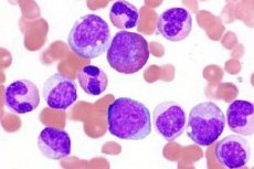
In size, cytoplasm color and nucleus shape, atypical cells are similar to lymphocytes and monocytes of peripheral blood. There are two types of virocytes: lymphocyte-like and monocyte-like, which differ in their size and cytoplasm composition. Mononuclears have a polymorphism of the nucleus shape with a spongy structure, the color of the cells from blue to pronounced basophilic. Many scientists consider them to be low-lymphocytes.
Atypical mononuclear cells in children
Virocytes can appear in the blood of patients of any age. Atypical mononuclear cells in children indicate a viral disease - mononucleosis. The disease occurs due to the Epstein-Barr virus, which affects parenchymatous organs and lymphoid tissue cells. The infectious process is localized in the pharynx, liver and spleen. Atypical cells appear with chickenpox, since the virus belongs to the same genus as the causative agent of mononucleosis. Its action reduces the protective properties of the immune system, opening the way for other pathologies.
Most often, atypical mononuclear cells are found in children aged 8-10 years. This is due to the fact that this age category is susceptible to many infectious diseases. Children under 1 year of age are the least likely to get sick; about 0.5% of all cases of mononuclear cells are found at this age. The infection is transmitted by airborne droplets, through contact between children, but it is unstable in the environment.
Symptoms of atypical mononuclear cells in children:
- Increased body temperature.
- Enlarged lymph nodes.
- Enlarged spleen/liver.
- Changes in the general composition of the blood.
- Plaque on the tonsils.
- Increased sweating.
In rare cases, petichial rashes (without a specific localization) and jaundice of the skin appear on the child's body. According to medical statistics, the virus is most often detected in boys, the peak incidence occurs in the autumn-winter and spring periods. Harmful microorganisms enter the mucous membrane of the upper respiratory tract and spread throughout the body, affecting the lymph nodes, liver and spleen. The incubation period lasts from 5 to 15 days.
To restore the normal level of virocytes in the blood, symptomatic and general strengthening therapy is carried out, which are aimed at eliminating the signs of infection. Antibiotics are not used, since they do not affect the virus. Vitamins of group B, C, P have medicinal properties.
 [ 8 ], [ 9 ], [ 10 ], [ 11 ], [ 12 ], [ 13 ], [ 14 ], [ 15 ]
Morphology of atypical mononuclear cells
The structure of viral cells largely determines the mechanism of their action and damage to the body. The morphology of atypical mononuclear cells indicates that their pathogen is the Epstein-Barr virus. These cells are herpes viruses, have a complex structure and contain DNA in the form of a double helix. The virus is resistant to low temperatures and drying.
The infection is transmitted by airborne droplets, contact and blood contact. The disease occurs in the form of sporadic outbreaks. As a rule, the infection is diagnosed in preschool and school-age children, mainly in boys. Children under one year do not get sick due to passive immunity. The disease is a seasonal phenomenon, exacerbation is observed in the winter-spring period. The disease does not recur, mortality is low, but there are data on isolated cases of spleen rupture, CNS damage and laryngeal stenosis.
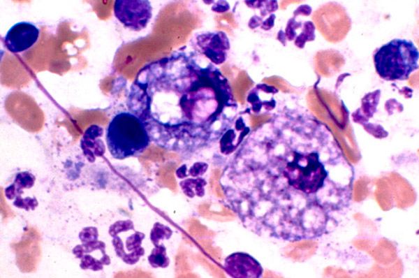
The virus penetrates through the mucous membrane of the upper respiratory tract and oropharynx. Adhesion occurs with the help of receptors located on the surface of epithelial cells. Virus reproduction leads to cell destruction, which causes the release of new generations of infection into the blood. Atypical mononuclear cells are infected B-lymphocytes with altered functional and morphological properties. Pathological changes in the immune system lead to the body being unable to completely neutralize the virus, which can remain latent in B-lymphocytes for life.
Cells morphologically similar to atypical mononuclear cells
Since virocytes indicate the presence of infection in the body, there are other cellular structures similar to them. Lymphocytes are cells morphologically similar to atypical mononuclear cells. They are similar in shape and size of the nucleus, cytoplasm. They are found in the blood in various viral diseases (rubella, flu, measles, chickenpox), autoimmune diseases, allergic reactions, vaccinations and various tumors.
Based on this, two types of atypical mononuclear cells are distinguished: monocyte-like and lymphocyte-like. Lymphocyte-like cells differ from lymphocytes in that they have foamy cytoplasm and are characterized by polymorphism of the nucleus with a spongy structure. That is, virocytes are modified T-lymphocytes. In rare cases, cells with granular a-naphthyl acetate esterase, not inhibited by NaF, are found. Virocytes have high activity of acid phosphatase, lactate, a-glycerophosphate and succinate dehydrogenases.
[ 16 ], [ 17 ], [ 18 ], [ 19 ]
Blood test for atypical mononuclear cells
Diagnostics of viral and infectious diseases involves various studies. Blood analysis for atypical mononuclear cells is performed for clinical symptoms of mononucleosis and similar diseases. Virocytes are determined using a general blood test, which evaluates the qualitative and quantitative cellular composition, leukocyte formula, plasma and cell volume ratio, color indices and ESR.
Altered T-lymphocytes are detected using the lymphocyte formula (the percentage ratio of different types of leukocytes). It is used to diagnose infectious, inflammatory and hematological diseases, as well as to assess the effectiveness of treatment. Mononucleosis is confirmed if more than 10% of atypical cellular structures are present in the blood.
[ 20 ], [ 21 ], [ 22 ], [ 23 ], [ 24 ]
Atypical mononuclear cells in mononucleosis
The presence of virocytes in the blood indicates an infectious disease caused by the Epstein-Barr virus. Atypical mononuclear cells in mononucleosis usually exceed the threshold of 10%. The incubation period is not precisely established, it varies from 5 to 21 days, and in some cases up to 1-2 months. The disease begins with a sharp increase in temperature, inflammation of the cervical lymph nodes, difficulty in nasal breathing. Later, an enlarged spleen and liver can be palpated in patients.
If mononucleosis has an atypical form, then the clinical symptoms are blurred, so the disease can only be recognized with the help of laboratory tests. Protracted and chronic forms are characterized by hematological changes and lymphadenopathy, which can persist for 4-6 months. The severity criterion is the severity of the intoxication syndrome, the duration of the disease and the presence of complications.
Atypical mononuclear cells in the blood require specific treatment. Glucocorticosteroids and vitamin complexes are used for therapy. Antibiotics are not used, since they do not affect the virus. If the liver is involved in the pathological process, the patient is prescribed diet table No. 5a / No. 5. In the absence of proper treatment, the disease has a poor prognosis and threatens complications. Most often, these are neurological disorders occurring as aseptic meningitis, encephalitis and other pathologies.
The norm of atypical mononuclear cells in the blood
To detect the first signs of any disease, a blood test is used. The norm of atypical mononuclear cells in the blood allows diagnosing various infectious and viral diseases. Normally, a healthy person should not have them. If their number exceeds the 10% barrier, this indicates infectious mononucleosis. Cells can be present in lymphocyte blood in healthy people, normally they contain no more than 1/6 of the total number of lymphocytes. The number of atypical mononuclear cells increases with:
- Autoimmune pathologies.
- Oncology.
- HIV infections.
- Infectious and viral lesions.
- Blood pathologies.
- Poisoning and long-term use of medications.
To determine the level of virocytes, a comprehensive blood test is performed with determination of the leukocyte formula. Normally, lymphocytes should be no more than 25-35%, and monocytes 2-6%. In pediatric patients, the number of cells is higher than in adults. A reduced level of atypical mononuclear cells and leukocytes is observed in:
- Liver failure.
- Use of cytostatics.
- Miliary tuberculosis.
- Aplastic anemia.
Their number fluctuates significantly in such pathologies as: blood diseases, poisoning, systemic collagenoses, acute infections. They are responsible for the body's immune response to various irritants. Virocytes are capable of differentiation, since they absorb damaged cells, harmful microorganisms and bacteria.
Causes of atypical mononuclear cells
In a healthy organism, with normal functioning of all organs and systems, virocytes are absent. The reasons for the appearance of atypical mononuclear cells are different. They are detected during laboratory examination, they are determined as a percentage of the number of leukocyte cells. If the concentration of atypical cells exceeds 10%, then the viral infection is in an acute or progressive form. In this case, additional blood tests and a doctor's consultation are carried out to clarify the diagnosis.
The main reason for the appearance of virocytes is infectious and viral diseases. Such illnesses are accompanied by fever, active enlargement of the liver, spleen and lymph nodes. Mononucleosis can change the quantitative composition of the blood. At the initial stage of the disease, along with an increase in the number of atypical cells, an increase in the number of band neutrophils is observed, and the number of segmented neutrophils decreases.