Medical expert of the article

New publications
Arachnodactyly
Last reviewed: 04.07.2025

All iLive content is medically reviewed or fact checked to ensure as much factual accuracy as possible.
We have strict sourcing guidelines and only link to reputable media sites, academic research institutions and, whenever possible, medically peer reviewed studies. Note that the numbers in parentheses ([1], [2], etc.) are clickable links to these studies.
If you feel that any of our content is inaccurate, out-of-date, or otherwise questionable, please select it and press Ctrl + Enter.
One of the rare hereditary connective tissue pathologies is arachnodactyly – a deformation of the fingers, accompanied by elongation of the tubular bones, skeletal curvatures, disorders of the cardiovascular system and organs of vision.
The disease was first described by Dr. Williams and the French pediatrician Marfan, who called it dolichostenomelia. Later, the term Marfan syndrome appeared in world medicine, and since 1902, the name arachnodactyly has been used. The pathology is related to congenital disorders of connective tissue, and one of the leading signs is considered to be elongated, thinned and curved "spider" fingers. [ 1 ]
Epidemiology
Arachnodactyly is considered a hereditary monogenic pathology of connective tissue, with autosomal dominant inheritance, high penetrance and varying degrees of expressivity.
In approximately 75-80% of cases the disease is hereditary, and in the remaining cases it occurs as a result of spontaneous mutations (in particular, due to point missense mutations). [ 2 ]
It has been proven that the pathogenetic features of arachnodactyly are caused by changes in several genes (in 95% of cases):
- in the fibrillin-1 gene (about one thousand mutations have been studied) on chromosome 15q21.1;
- the TGFβR1 or TGFβR2 gene on chromosome 9 and 3p24.2-P25.
These changes directly affect the extent of clinical manifestations.
Mutations in the α-2 chain of type I collagen have been described in 5% of patients.
The prevalence of arachnodactyly is about 2 cases per 5 thousand people. There is no racial or gender determination. [ 3 ]
Causes arachnodactyly
Arachnodactyly occurs as a result of spontaneous mutation. The disease is not characterized by the generally known signs of congenital hereditary pathologies, and elongated fingers are manifested mainly in Marfan syndrome and homocystinuria.
The disease is rare, and all patients found had a mutation of the FBN1 gene (fibrillin gene) located on chromosome 15 in the q21.1 region. The clinical polymorphism of the syndrome can be explained by the mass of mutations, with about 15% being mutational changes that occurred during conception. [ 4 ]
Atypical varieties of arachnodactyly are caused by mutations in other genes: a relationship has been discovered between the development of the pathology and mutations in the LTBP3, R gene, which is localized on chromosome 14 in the q24 region and transforms growth factor.
The overwhelming majority of mutations are so-called missense mutations (approximately 57%). More than 20% are small deletions, with loss of chromosomal parts, about 12% are splicing site mutations, 8% are nonsense mutations, and 2% are large-scale rearrangements and deletions. [ 5 ]
Congenital contractural arachnodactyly is caused by a disorder in the synthesis of the protein agent fibrillin-2. The problem is caused by a mutation in the FBN2 gene, located on the fifth chromosome in the q23-q31 region. All mutations are predominantly point mutations, switching the codon to code for another amino acid. [ 6 ]
Homocystinuria develops against the background of impaired metabolism of the essential amino acid methionine. The basis of the pathology may be the loss of activity or its decrease in the following enzyme substances:
- Cystathionine beta synthase (CBS) enzyme. The pathology is B 6 resistant and manifests itself with signs characteristic of the B 6 -independent form, however, the introduction of a B 6 -vitamin preparation does not lead to improvement.
- Enzyme N5, N10 methylenetetrahydrofolate reductase (MTHFR). This enzymatic agent is a substrate that causes the demethylation of homocysteine and its transformation into methionine, an essential amino acid that is a component of many protein and peptide substances.
- Enzyme N5 methylenetetrahydrofolate. We are talking about B 6 dependent form of pathology, which develops with deficiency of vitamin B 6. Introduction of vitamin preparation into the body allows to improve the patient's condition.
- Homocysteine transmethylase enzyme. Develops against the background of metabolic disorders of cobalamin – vitamin B 12.
Risk factors
Arachnodactyly is a genetically determined disease characterized by systemic damage to connective tissue. The pathology is associated with changes in the fibrillin 1 gene, localized on the short arm of chromosome 15 in locus 21.1.
Arachnodactyly is inherited in an autosomal dominant manner, with high penetrance and variable expressivity. The pathology can manifest itself on both the male and female side.
Pathogenesis
More than 50% of human body mass is made up of connective tissue structures, in particular the skeleton, skin, vascular network, lymph and blood.
Connective tissue cells are represented by fibroblasts and their subtypes: osteoblasts, chondrocytes, keratoblasts, odontoblasts, as well as macrophages and mast cells.
Embryonic connective tissue is the material for the formation of constitutional, genetic and epigenetic components. Connective tissue diseases are reflected to one degree or another on the entire body as a whole, on its functional capacity and constitution.
In arachnodactyly, there is a substitution of nucleotides in the gene that contains structural information about the fibrillin-1 peptide. This protein substance belongs to the glycoprotein group, participates in the microfibrillar system and forms the basis of elastic fibrils of connective tissue.
The tissue maintains a stable structure due to the intercellular matrix, which contains many growth factors that ensure systematic cellular renewal. Large vessels and ligaments contain numerous elastin fibrils, the damage to which causes the main symptoms of arachnodactyly.
The transforming growth factor β is significantly damaged: its inactive form is not bound, which leads to an increase in the biological activity of this factor.
Fibrillary disorders lead to abnormal formation of fibers, resulting in loss of strength and elasticity of the skin and other connective tissue structures.
When the collagen structure is disrupted, the primary link in hemostasis is disrupted.
Experts note the involvement of hormonal imbalances in the emergence and progression of anomalies in connective tissue structures, including arachnodactyly. Thrombotic symptoms are determined by rheological disorders - in particular, changes in blood viscosity in the modified vessels of the brachiocephalic region.
The digestive tract is affected due to collagen disorders: hypomotor biliary dyskinesia, hernias of the esophageal opening of the diaphragm, hepatobiliary anomalies, chronic form of “erased” gastroduodenitis, dolichosigma are observed.
How is arachnodactyly inherited?
Arachnodactyly is inherited as an autosomal dominant trait, but can also occur as a result of spontaneous mutations. That is, the pathology does not necessarily have to manifest itself in one of the close relatives - for example, in parents, grandparents. Mutations underlying the pathology can make themselves known randomly.
Symptoms arachnodactyly
In medicine, a distinction is made between latent and overt arachnodactyly. In latent arachnodactyly, changes are present, but they are isolated and limited to one or two organ systems. Overt arachnodactyly manifests itself in multiple visible disorders with varying intensity of symptoms. In this case, the patient's condition may be relatively stable for decades, or steadily progress, manifesting itself in relation to other organs and systems.
Often, arachnodactyly syndrome can be detected within the first few days after the baby’s birth.
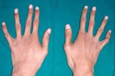
The main external sign of arachnodactyly is thin and elongated fingers, which, due to short tendons, acquire a typical curvature and resemble spider legs.
Spider fingers or arachnodactyly become clearly visible almost from the newborn period. But the sign becomes more obvious at about three years of age.
Growth indicators are also characteristic: children are usually tall, forearms and shins are long, limbs are disproportionate and thin. The disproportion is especially obvious in the high localization of the waist and kneecaps. [ 7 ]
Other skeletal disorders include:
- narrowed and elongated skull shape (dolichocephalic) with an abnormally formed facial area;
- funnel-shaped configuration of the chest (the so-called "bird");
- curved spine;
- congenital hip dislocations;
- habitual knee-shoulder dislocations;
- curvature of the heel bone;
- osteophyte formations – bone growths;
- flat feet;
- insufficient development of the fat layer.
Additional signs of arachnodactyly may include:
- myopia;
- bluish sclera;
- subluxation of the lens;
- disorders of the heart and blood vessels (heart defects, aortic aneurysm);
- pronounced thinness;
- joint hypermobility;
- "arched" sky.
Arachnodactyly is characterized by elongated tubular bones, causing various skeletal curvatures. In addition, many patients have pyramidal disorders, asymmetry of tendon reflexes, anosocoria and nystagmus against the background of normal mental development.
If we are talking about arachnodactyly, accompanying homocystinuria, then the patient may experience the following symptoms:
- periodic attacks of convulsions;
- secondary glaucoma with lens subluxation;
- retinal detachment, astigmatism;
- damage to arterial trunks (including kidneys, brain);
- osteoporosis;
- thrombosis;
- mental retardation.
Arachnodactyly in newborns
In most cases, arachnodactyly can be identified in a child within the first few days of his life. However, with age, the symptoms tend to worsen.
The first signs that allow one to suspect the disease are usually the following:
- unusually long limbs and fingers;
- underweight in an adequate physical condition of the child;
- elongated, long face;
- lack of body fat, poorly developed muscles;
- excessive joint flexibility.
From about four years of age, against the background of arachnodactyly, the chest begins to change, the spinal column becomes curved, and flat feet develop.
In terms of the visual organs, myopia, ectopia of the lens, altered configuration of the cornea, strabismus, hypoplastic processes in the iris and retina are observed. Such disorders can be noticed already in the first 2-3 years of a child's life, although they progress over the years.
The greatest danger is posed by disorders of the cardiovascular system. If such disorders are present, then in the absence of appropriate treatment there are high risks of death of the patient already in childhood. Among the especially dangerous pathologies, one can single out damage to the vascular walls, cardiac malformations, defects of the coronary vessels. Sometimes, in children of the first year of life, increasing cardiac insufficiency is already detected.
Arachnodactyly may be accompanied by damage to other organs, in particular, the nervous system, bronchi and lungs, skin, and genitourinary system. Such disorders are detected during diagnostic procedures.
Complications and consequences
The most common adverse effects of arachnodactyly are damage to the heart and respiratory organs. In addition, problems with vision appear, up to and including its loss.
Patients with arachnodactyly require complex therapy of all existing pathologies. If such treatment is not carried out, the life expectancy of patients is significantly reduced: rare patients without medical intervention manage to live to the age of 40. Over the years, problems with the cardiovascular system worsen, the risk of extensive hemorrhages with a tragic outcome increases.
People suffering from arachnodactyly should undergo regular examinations and treat concomitant pathologies: only under this condition do they have every chance of living a long life without serious complications.
Marfan syndrome and arachnodactyly require special attention if the female patient becomes pregnant. In such a situation, it is important to regularly and thoroughly examine the patient, and to medically support the condition of her cardiovascular system. This is the only way to guarantee a successful pregnancy and a safe birth of the child.
To avoid adverse effects, patients with arachnodactyly should be examined regularly by doctors, prevent the development of cardiovascular and musculoskeletal pathologies. They are recommended to lead an exclusively healthy lifestyle, with moderate physical activity.
Diagnostics arachnodactyly
Clinical diagnostics for arachnodactyly includes collecting complaints, family history, phenotype assessment, as well as anthropometric and physical examinations. It is important to determine the presence of manifestations of connective tissue pathologies in direct relatives: for this purpose, a genealogical study is carried out. [ 8 ]
Laboratory diagnostics for arachnodactyly involves studying the condition of certain types of connective tissue, represented by bone and cartilage tissue, blood, and lymph. The most informative is considered to be a study of the levels of oxyproline and glycosaminoglycans in the daily volume of urine. In addition, the content of lysine, proline, and oxyproline in the blood is assessed. To determine altered ratios of different types of collagens and structural abnormalities of collagen fibers, typing is prescribed by the method of indirect immunofluorescence using polyclonal antibodies to collagen and fibronectin. [ 9 ] Depending on the indications, the following tests are performed to reflect the quality of connective tissue metabolic processes:
- glycosaminoglycans, fibrillin, fibronectin;
- study of hydroxyproline, markers of type 1 collagen biosynthesis, aminoterminal propeptides of type 1 procollagen, markers of type 1 collagen degradation, galactosyloxylysine, deoxypyridinoline;
- analysis of collagen metabolism regulation (study of matrix metalloproteinases and tissue inhibitors of matrix metalloproteinases, transforming growth factor);
- analysis of micro and macroelements (content of calcium and magnesium, phosphorus, iron, sulfur and copper, cobalt and selenium, zinc and manganese, fluorine and vanadium, silicon and boron);
- study of markers of bone tissue formation and the rate of remodeling processes (analysis of the content of osteocalcin, bone alkaline phosphatase, parathyroid hormone, somatotropic hormone, prolactin, vitamin D3 , pentosidine, homocysteine in urine and blood).
Instrumental diagnostics aims to identify existing developmental defects and assess the functional state of organs and systems. Cardiovascular disorders in arachnodactyly require mandatory inclusion in the diagnostic list of such procedures as:
- electrocardiography;
- 24-hour ECG monitoring;
- ultrasound examination of the heart and blood vessels.
Vascular anomalies are examined using computed tomography or magnetic resonance imaging. Additional diagnostic procedures may include:
- Ultrasound of abdominal organs, genitourinary system;
- X-ray of the chest organs, hip joints; [ 10 ]
- CT or MRI of the spine.
After all necessary tests, the patient is referred for genetic counseling.
Differential diagnosis
There is a phenotypic similarity between Beals syndrome and arachnodactyly in Marfan syndrome (mutations of the FBN2 and FBN1 genes, respectively), which is due to the almost complete identity of the protein substances fibrillin 1 and fibrillin 2. It is not surprising that the second name of Beals syndrome is contracture arachnodactyly. [ 11 ]
Differentiation is also carried out with other connective tissue pathologies:
- Stickler syndrome;
- Ehlers-Danlos syndrome;
- homocystinuria;
- arthrogryposis.
Beals syndrome is characterized by a marfanoid phenotype, congenital flexion contractures of large and small joints, wrist and foot arachnodactyly, curvature of the spine, and abnormal ear shape (so-called "crumpled" auricles). Molecular analysis of the FBN2 gene is performed to confirm the diagnosis.
Arthrogryposis is characterized by multiple joint contractures, short stature, and decreased intelligence. The shape of the fingers and ears is unremarkable. [ 12 ]
Ehlers-Danlos syndrome is characterized by kyphoscoliosis, funnel-shaped curvature of the chest, pronounced flat feet, and impaired refraction. Increased mobility and joint dislocations, high skin extensibility and sensitivity are typical.
Stickler syndrome is characterized by changes in joint volume and stiffness.
Who to contact?
Treatment arachnodactyly
Arachnodactyly is a genetic pathology, and currently medicine does not have methods for correcting gene defects. Therefore, treatment is aimed at optimizing the patient's condition, preventing the aggravation of the pathology, and eliminating symptomatic manifestations. Complex therapy is prescribed, involving several medical specialists at once, depending on the type of the most pronounced symptoms: often it is necessary to additionally contact an orthopedist, cardiologist, ophthalmologist, gastroenterologist, and doctors of other specialties. [ 13 ]
Among the recommendations for clinical management of patients, the following are considered general principles:
- limiting physical activity, but not completely eliminating it (as part of supporting the cardiovascular system);
- drug therapy;
- if necessary – surgical correction of the most affected areas of the heart and blood vessels;
- orthopedic correction;
- spa treatment, physiotherapy, therapeutic exercise.
The diet of patients with arachnodactyly should include a sufficient amount of high-protein products enriched with microelements, vitamins, and fatty acids. It is recommended to eat meat, fish, seafood, beans, and nuts. If arachnodactyly is caused by homocystinuria, then the consumption of animal protein is sharply limited.
Children with a thin build and tall stature are advised to introduce cottonseed and soybean oil, sunflower seeds, pork fat and lard into their diet from an early age. Additionally, they offer preparations containing polyunsaturated fatty acids such as Omega, which inhibit the production of somatotropic hormone.
To normalize protein metabolism, vitamins of the B group are prescribed. They can also be obtained from food products: buckwheat, peas, liver.
It is very important that ascorbic acid enters the patient's body with food. For this purpose, rosehip infusion, bell peppers, cabbage, citrus fruits, as well as sea buckthorn and leeks are necessarily included in the diet.
If necessary, patients are offered orthopedic correction, which is necessary to reduce the load on the spine and joints. For this purpose, orthopedic shoes, knee and other devices, insoles, elastic bandages are used.
Surgical treatment is performed according to indications.
Medicines
Drug treatment for arachnodactyly is carried out 1-2 times a year, depending on the patient's condition and the severity of pathological symptoms. The duration of the treatment course is determined individually and is on average 4 months. [ 14 ]
To stimulate collagen formation, the following drugs are prescribed: Piaskledin 300, L-lysine or L-proline in combination with complex multivitamin preparations containing ascorbic acid, B vitamins, tocopherol, magnesium, zinc, selenium, copper.
Among chondroprotectors, the most optimal use is chondroitin sulfate and glucosamine sulfate - drugs that participate in the regulation of chondrocyte metabolism, suppression of enzyme synthesis, increase in the sensitivity of chondrocytes to enzymatic action, stimulation of anabilistic processes, etc. Combined agents are considered optimal such drugs: Teraflex, Arthroflex, Arthra, etc.
Mineral metabolism is stimulated by drugs that normalize phosphorus-calcium processes. The drugs of choice are often active forms of vitamin D: Alpha D 3 - Teva, Oxidevit, Bonviva, etc. At the same time, phosphorus, calcium, and magnesium preparations are used. During treatment, the level of calcium and phosphorus in the blood or urine is checked approximately once every 20 days, and a blood test for alkaline phosphatase is performed.
To improve the bioenergetic state of the body, it is possible to prescribe Phosphaden, Riboxin, Lecithin, Elkar, Coenzyme Q10.
An approximate treatment regimen might look like this:
- Chondroprotector within the age-appropriate dose, taken with food and sufficient amount of water. Duration of one treatment course is 3-4 months.
- L-proline in a dosage of 500 mg (for children from 12 years and adults) is taken half an hour before meals, 1-2 times a day, for six weeks. If indicated, an amino acid complex can be additionally prescribed - L-proline, L-lysine, L-leucine in the amount of 10-12 mg / kilogram of weight. The intake is carried out 1-2 times a day, for two months.
- Vitamin and mineral complex preparations Centrum, or Vitrum, or Unicap, in dosage taking into account age. Duration of administration is 4 weeks.
This treatment regimen is appropriate if a patient with arachnodactyly complains of problems with the musculoskeletal system, and test results indicate increased excretion of glycosaminoglycans in a daily urine test and decreased levels of free amino acids in the blood.
As a rule, the treatment is well received by patients, without any particular side effects. If hypersensitivity reactions are detected, the drugs are replaced and the treatment regimen is adjusted.
Physiotherapy treatment
Physiotherapeutic procedures for arachnodactyly are prescribed according to indications. For example, in case of insufficient osteogenesis, to improve the healing of bone fractures, or in case of signs of osteoporosis, electrophoresis with 5% calcium chloride, 4% magnesium sulfate, 2% copper sulfate or 2% zinc sulfate is recommended.
If vegetative-vascular disorders are detected, 1% sodium caffeine benzoate, ephedrine hydrochloride or mesaton are used.
To activate the functioning of the adrenal cortex, drug electrophoresis with 1.5% ethimizole and decimeter therapy to the adrenal region are prescribed. [ 15 ]
To stabilize the tone of blood vessels, water procedures are recommended that promote vascular "gymnastics". Baths such as carbon dioxide, pine, chloride-hydrogen, hydrogen sulfide, and radon are excellent. Rubdowns, dousings, contrast showers, foam and salt baths are practiced.
To improve the condition of cartilage tissue, magnetic therapy, inductotherapy, laser therapy, and electrophoresis with dimethyl sulfoxide (Dimexide) are used.
Surgical treatment
Surgical operations for arachnodactyly are prescribed strictly according to indications. For example, if severe hemodynamic disturbances are detected against the background of prolapse of valve cusps, massive aortic aneurysm, valve replacement and damaged segment of the aorta are performed. [ 16 ]
If there are pronounced functional disorders of the respiratory and cardiovascular systems that arise as a result of severe curvature of the chest, thoracoplasty is performed.
In case of progressive pain syndrome caused by severe scoliosis of 3-4 degrees, surgical intervention is also indicated. From the ophthalmological point of view, absolute indications for lens removal are subluxation of the lens complicated by secondary glaucoma, as well as cataracts and degenerative changes in the retina with a high risk of detachment.
Any operation on patients with arachnodactyly and other disorders affecting connective tissue structures is performed only at the stage of relative clinical and biochemical relief. After the intervention, long-term medical observation and intensive therapy with drugs that improve connective tissue metabolism are mandatory.
Prevention
Arachnodactyly is a rare genetic pathology. Sometimes it occurs spontaneously in apparently healthy parents. Therefore, it is very, very difficult to prevent the disease in advance.
If one of the potential parents has a burdened family history - that is, it is known that someone in the family suffered from arachnodactyly - then the spouses should visit a geneticist and undergo a genetic examination at the stage of planning a child. After confirming the pregnancy, the doctor performs prenatal diagnostics, including an ultrasound examination of the fetus and a number of biochemical tests:
- maternal blood test;
- amniotic fluid analysis;
- chorionic villus sampling;
- analysis of placental cells and umbilical cord blood.
Patients with arachnodactyly are under medical monitoring throughout their lives. Preventive measures in patients are aimed at preventing complications. For this purpose, cardiac surgical correction is performed, drug treatment is prescribed to eliminate the risk of thrombus formation. Antibiotic therapy courses are periodically administered.
Forecast
The prognosis for the life of patients with arachnodactyly is primarily determined by the severity of concomitant disorders - in particular, diseases of the cardiovascular, musculoskeletal system and visual organs. Most often, the pathology is complicated by rupture and dissection of the aorta. This must be taken into account and timely cardiac surgery correction must be performed, which will help prolong the patient's life and improve its quality.
If the diagnosis was made in a timely manner, then if the doctor's recommendations are followed, the prognosis can be called conditionally favorable. Medical support significantly alleviates the course of arachnodactyly, and patients have the opportunity to live a normal and relatively full life without developing serious complications.