Medical expert of the article

New publications
Aper syndrome
Last reviewed: 04.07.2025

All iLive content is medically reviewed or fact checked to ensure as much factual accuracy as possible.
We have strict sourcing guidelines and only link to reputable media sites, academic research institutions and, whenever possible, medically peer reviewed studies. Note that the numbers in parentheses ([1], [2], etc.) are clickable links to these studies.
If you feel that any of our content is inaccurate, out-of-date, or otherwise questionable, please select it and press Ctrl + Enter.
 [
[ Causes Aper syndrome
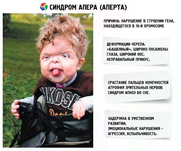
The development of the syndrome is provoked by a disorder in the structure of a gene located on chromosome 10. This gene is responsible for the process of finger separation, and in addition, for the timely closure of cranial sutures.
In addition, infectious diseases suffered during pregnancy (such as rubella or tuberculosis, syphilis or flu, as well as meningitis), and exposure of the mother to X-rays are considered to be the causes of the pathology. Most often, such a syndrome is detected in children born to elderly parents.
Pathogenesis
Apert syndrome is hereditary. Its type is autosomal dominant (i.e., in a family in which one of the future parents is ill, the probability of having a baby with this disease is 50-100%).
A unique mutation in fibroblast growth factor receptor 2 (FGFR2) results in an increased number of progenitor cells that develop along the osteogenic pathway. This ultimately results in increased subperiosteal bone matrix formation and premature ossification of the cranial sutures during fetal development. The order and rate of suture melting determines the degree of deformity and disability. Once the suture material has healed, growth of other tissues perpendicular to this suture is limited, and the fused bones act as a single bone scaffold.
The first genetic evidence that syndactyly in Apert syndrome is due to a defect in the keratinocyte growth factor receptor (KGFR) was the observation of a correlation between KGFR expression in fibroblasts and the severity of syndactyly.
Amblyopia and strabismus are more common in patients with the FGFR2 Ser252Trp mutation, and optic disc atrophy is more common in patients with the FGFR2 Pro253Arg mutation. Patients with FGR2 Ser252Trp mutations have a significantly higher prevalence of visual impairment compared to patients with the FGFR2 Pro253Arg mutation.
Symptoms Aper syndrome
Some manifestations of the disease are clearly noticeable already at the birth of the baby, because they develop inside the mother's womb. Among the main symptoms of the syndrome:
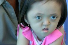
- The skull is deformed - it stretches in height, acquiring a "tower" appearance; in addition, the eyes are set wide and slightly bulging (because the size of the eye sockets decreases); the nose widens and an incorrect bite is formed (the upper teeth protrude excessively);
- The fingers of the limbs are completely fused (mainly the ring finger, as well as the middle finger and index finger) and look like a skin membrane or a complete fusion of bones; in addition, extra fingers may grow;
- Mental retardation (does not occur in everyone);
- The optic nerves atrophy, as a result of which visual acuity decreases (in some cases, complete loss of vision develops);
- Increased intracranial pressure, which occurs as a result of premature closure of cranial sutures, manifests itself in the form of headaches and vomiting with nausea;
- Since the upper jaw remains underdeveloped, breathing problems arise;
- Sleep apnea syndrome is a common occurrence.
- Emotional manifestations – aggression, lack of restraint, strong irascibility.
Diagnostics Aper syndrome
To make a diagnosis, the following steps are required:
- The doctor should analyze the patient's complaints, as well as the medical history. It is necessary to find out whether there were cases of similar pathology in the family;
- Neurological examination to assess the shape of the skull and the patient's intellectual development (special questionnaires as well as interview);
- Examination of the fundus to detect the presence of symptoms of increased intracranial pressure (swelling of the optic disc, as well as blurring of its edges);
- To assess the condition of the skull, an X-ray is performed;
- Computer and magnetic resonance imaging of the head to examine the structure of the brain and skull layer by layer, to determine the presence of symptoms of premature fusion of cranial sutures, and in addition, hydrocephalus (due to increased intracranial pressure, excess cerebrospinal fluid accumulates (this is the cerebrospinal fluid that promotes the metabolic process, as well as nutrition of the brain));
- X-ray of the feet and hands to determine the cause of the fusion of the fingers (this is important for planning subsequent surgical intervention);
- A consultation with a medical geneticist and a neurosurgeon may be prescribed.
Tests
Genetic analysis of common mutations occurring in the FGFR2 type gene is being conducted.
Differential diagnosis
This syndrome must be differentiated from other genetic pathologies in which craniosynostosis is observed. These are diseases such as Pfeiffer, Crouzon, Saethre-Chotzen and Carpenter syndromes. Molecular genetic testing methods are used to exclude these anomalies.
Who to contact?
Treatment Aper syndrome
Surgery is considered the only effective treatment for Apert syndrome – it helps correct individual physical defects and also corrects mental retardation.
During this procedure, the coronal suture is closed to prevent possible brain injury. The most common technique is craniofacial distraction, which involves graded skull traction. Orthodontic and/or orthognathic surgery is performed to remove individual facial defects.
In addition, patients undergo surgical correction of finger fusion.
References
Medical Genetics. National Guide. Edited by Ginter E.K., Puzyrev V.P. GEOTAR-Media. 2022