
Medical expert of the article

New publications
Angiofibroma in children and adults: causes, treatment
Last reviewed: 04.07.2025

All iLive content is medically reviewed or fact checked to ensure as much factual accuracy as possible.
We have strict sourcing guidelines and only link to reputable media sites, academic research institutions and, whenever possible, medically peer reviewed studies. Note that the numbers in parentheses ([1], [2], etc.) are clickable links to these studies.
If you feel that any of our content is inaccurate, out-of-date, or otherwise questionable, please select it and press Ctrl + Enter.
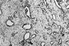
The term "angiofibroma" is used for any tumors that can be classified as angiomas with the presence of connective tissue fibers in them, or as fibromas penetrated by a vascular network. Such neoplasms include, for example, fibrous papules, sebaceous adenomas, nail fibromas, pearly papules, Koenen's tumors, etc.
Epidemiology
- Angiofibroma is diagnosed relatively frequently.
- The tumor can develop regardless of age, but it is more often found in patients over 40 years of age.
- Angiofibroma occurs regardless of race.
- The spread of the neoplasm is the same in both male and female patients. However, in adolescence, boys are more likely to get sick.
Causes angiofibromas
Experts cannot name the exact cause of the development of a disease such as angiofibroma. There are several theories that are used to explain the causes of the pathology:
- Hormonal theory.
Frequent diagnosis of the disease in children in adolescence has suggested that a certain risk factor may be a hormonal imbalance. However, information regarding this assumption is contradictory: some scientists do not admit the possibility of a negative impact of hormonal imbalance, while others confirm the involvement of the function of the sex glands. Experts have not yet made a final conclusion.
- Genetic theory.
This assumption is considered one of the most common. Information about the complete or partial absence or disruption of Y and X chromosomes in the cells of the neoplasm serves as confirmation. Such conclusions are not final and require additional research.
- Age influence theory.
Some specialists tend to view the disease as a result of age-related changes in the body.
In addition, there are suggestions that certain factors influence the development of the disease.
Risk factors for developing angiofibroma may include the following:
- injuries to the face, head, nose, etc.;
- inflammatory diseases, especially chronic inflammations (for example, chronic sinusitis, etc.);
- the presence of harmful substances, poor ecology, unacceptable lifestyle, etc.
Pathogenesis
Angiofibroma can occur at almost any age, in people of any gender and race.
The most common neoplasm of the nasopharynx: angiofibroma develops on the basis of the pharyngeal main fascia and is a basal or basosphenoidal type of tumor. The growth covers the surface of the sphenoid bone and/or the area of the posterior cells of the ethmoid bone (sphenoethmoidal type).
In some cases, the disease begins in the pterygopalatine fossa, spreading into the nasal cavity and into the space behind the jaw. This type of angiofibroma growth is diagnosed with the pterygomaxillary variant of tumor development.
This neoplasm is considered benign, although it often grows rapidly, affecting nearby tissues, cavities, and sinuses. Angiofibroma that has grown into the cranial cavity is especially dangerous.
Symptoms angiofibromas
Angiofibroma looks like a small (3-15 mm) single nodule with clearly defined contours and an elastic structure. The color of the formation can vary from pinkish-yellow to brown.
The nodule is usually slightly raised above the surrounding tissues.
In some cases, the nodule has a rich capillary network and has a translucent structure: this can be seen with a microscope.
The tumor is found in most cases on the mucous tissues of the upper respiratory tract, or on the upper or lower extremities, less often on organs (for example, kidneys).
The first signs of a neoplasm depend on its location. For example, if the nasopharynx is affected, the patient complains of difficulty breathing through the nose, up to its complete absence. Congestion may occur only on one side, gradually worsening. However, no nasal drops will relieve this problem.
Over time, snoring (even when awake), a feeling of dryness in the throat, and one- or two-sided hearing loss appear. Frequent headaches and spontaneous nosebleeds are possible.
Angiofibroma at the initial stage does not always manifest itself with changes in the blood picture. In most patients, signs of anemia - a decrease in the number of red blood cells and hemoglobin, are detected already at later pathological stages.
Angiofibroma in a child may resemble the signs of adenoids, therefore, in childhood it is customary to differentiate these pathologies.
In children, non-ossifying fibromas and metaphyseal defects are the most frequently diagnosed skeletal pathologies; these are relatively common skeletal pathologies in pediatric patients.
As the tumor grows, the patient's health deteriorates. Sleep becomes restless, appetite is disturbed, facial features change (deformation and asymmetry are possible).
Stages
Juvenile angiofibroma is conventionally divided into several stages:
- The neoplasm has limited growth and does not extend beyond the nasal cavity.
- The neoplasm grows into the pterygopalatine fossa or nasal sinuses.
- The tumor extends into the orbit or infratemporal fossa without intracranial growth (stage ) or with extradural growth (stage ).
- The tumor is characterized by intradural growth without involving the cavernous sinus, pituitary gland, or optic chiasm (stage ), or with involvement of the listed areas (stage ).
Forms
The species classification of the disease is associated with the localization of the tumor, as well as with some of its structural and pathogenetic features.
- Angiofibroma of the nasopharynx is a benign formation that consists of vascular and connective tissue and develops in the nasopharyngeal cavity. Most often, such a disease as angiofibroma of the nose is found in adolescent boys, so it is also called the term "juvenile angiofibroma" this is the most common neoplasm in pediatric patients.
- Angiofibroma of the skin is often mistaken for hemangioma: it is a connective tissue formation that looks like a wart. Such a tumor is usually round, has a base and penetrates deep into the skin. It is most often found on the upper and lower extremities, as well as in the neck and face. Mostly women aged 30-40 are ill.
- Laryngeal angiofibroma is a benign tumor that occupies the vocal cords. A similar term for the disease is vocal cord angiofibroma. The tumor has a reddish or bluish tint, has an uneven structure and is located on a stalk. The first signs of the disease are the appearance of characteristic hoarseness up to complete loss of voice.
- Angiofibromas of the face are located in different areas. They can be detected at any age. The main clinical symptom of the disease is the sudden appearance of a small, dense or elastic growth. There are usually no other symptoms. If the tumor is constantly touched and damaged, it can bleed and rapidly increase. Often, the formation is found in the nasal or ear cavities, on the eyelids.
- Angiofibromas of the face in tuberous sclerosis are typical manifestations of this pathology. Tuberous sclerosis is a hereditary neuroectodermal disease, the main symptoms of which are seizures, mental retardation, and the appearance of neoplasms such as angiofibromas. Neoplasms are found in more than half of patients suffering from tuberous sclerosis. They can be detected after the age of 4.
- Angiofibroma of the skull base is the rarest and, at the same time, the most severe form of the disease, in which a benign formation is formed in the bone structure of the skull base. The disease is difficult to diagnose at an early stage, due to its similarity to hypertrophic and inflammatory pathologies of the nasal and pharyngeal region. Angiofibroma of this type tends to grow rapidly with the destruction of cranial bones and spread to nearby anatomical brain structures. The overwhelming majority of patients are boys and young people aged 7-25 years.
- Angiofibroma of soft tissues often develops in patients on the skin, mammary glands, tendons. Soft tissues on the limbs, in the torso, face or neck are mainly affected. In some cases, tumor processes also develop in the area of internal organs - in the uterus, ovaries, lungs, mammary glands.
- Renal angiofibroma is a benign formation that can last for a long time without showing any symptoms. Rarely, the disease is detected due to the appearance of pain in the kidney. The disease is cured surgically: if the neoplasm is small, dynamic monitoring of the tumor is possible.
Complications and consequences
In itself, a neoplasm such as angiofibroma belongs to the benign group, and only in extremely rare cases can the disease become malignant.
However, tumor development often occurs rapidly. Rapid growth of the tumor can provoke destruction of nearby structures: even dense tissues, such as bones, are damaged. Therefore, rapid tumor growth can cause prolonged and massive bleeding (often repeated), deformation of the facial part, disorders of respiratory and visual function. In order to prevent complications of angiofibroma, it is very important to promptly determine the presence of a neoplasm and begin treatment.
Diagnostics angiofibromas
The diagnosis of the disease has its own characteristics. First of all, during the examination the doctor will pay attention to the number of pathological formations, their nature. The patient will be asked questions about the presence of similar pathologies in family members and relatives, about the detection of malignant diseases in close people, about possible disorders of the central nervous system.
If multiple tumor foci are detected, the patient is recommended to undergo examination for tuberous sclerosis or MEN I.
Blood tests will indicate the presence of anemia and inflammation in the body. Thus, a decrease in hemoglobin levels to 80 g / liter and red blood cells to 2.4 per 10¹² / l may be observed. Blood biochemistry often shows a decrease in total protein, albumin, as well as an increase in the amount of ALT, AST and alkaline phosphatase.
A tissue biopsy is a study of a portion of tissue using a microscope, which will allow one to accurately determine the benign nature of the neoplasm.
In addition, the doctor often prescribes a blood test for specific tumor markers to rule out malignancy.
Additional instrumental diagnostics often include fibroscopy or endoscopy. Such procedures are performed using a special device - an endoscope, which will allow you to detect and examine a tumor in cavities, for example, in the nasopharynx. This method will help assess the superficial state of the tumor, visualize the vascular network, and determine the presence of an inflammatory reaction.
Unfortunately, the radiographic method is uninformative in this situation. Using this method, you can see the presence of a formation, but you will not be able to conduct a differential diagnosis of angiofibroma.
Differential diagnosis
Differential diagnosis is carried out with syringoma, angioleiomyoma, cavernous hemangioma, flat pigmented nevus, Osler-Rendu disease, angiokeratoma, squamous cell carcinoma, etc.
Who to contact?
Treatment angiofibromas
In the vast majority of cases, patients with angiofibromas are prescribed surgical treatment. The volume and type of surgical intervention is selected taking into account the stage and localization of the pathological focus.
If the tumor process has a significant spread and is penetrated by a large number of vessels, then it may be necessary to pre-prepare the patient. This is done to reduce the risk of bleeding during and after the operation.
Treatment may include the following methods:
- X-ray endovascular occlusion – used when it is impossible to perform a full surgical intervention, as an initial stage of radiation therapy. The use of the method allows to reduce the volume of intraoperative blood loss.
- Radiation therapy – helps in about 50% of cases of angiofibroma, but can be accompanied by a large number of complications. For this reason, it is used only when it is impossible to perform a full surgical intervention.
Medicines
Drug treatment is aimed at eliminating the main painful symptoms of angiofibroma, as well as alleviating and prolonging the life of patients.
- If pain occurs, it is recommended to take Baralgin or No-shpa 1-2 tablets up to three times a day. Duration of treatment is from one to four weeks.
- To improve organ function and strengthen blood vessels, Stimol is prescribed in the amount of 1 packet twice a day, as well as multivitamin complexes - for example, Duovit, 2 tablets daily for 4 weeks, or Vitrum, 1 tablet daily for 4-12 weeks.
Combination chemotherapy is used on an individual basis as needed. The following may be offered:
- combination of Adriamycin, Sarcolysine and Vincristine;
- combination of Vincristine, Decarbazine, Adriamycin, Cyclophosphamide.
For example, such treatment can be used before and/or after surgery.
Vitamins
High-quality treatment of angiofibroma is impossible to imagine without vitamin therapy. Eating certain vitamins does not affect tumor growth, but it helps strengthen blood vessels, prevent anemia, and normalize the immune system.
- Vitamin A will prevent secondary infection, improve tissue turgor and speed up recovery. This vitamin can be obtained by eating plant foods or by purchasing capsules with an oily vitamin solution in pharmacies.
- Tocopherol prevents the formation of blood clots in blood vessels, rejuvenates and renews tissues. The drug is available in any pharmacy without a prescription, and is also present in products such as butter and sunflower oil, nuts, seeds and milk.
- Ascorbic acid takes part in the regulation of recovery reactions in the body, normalizes the production of hormones. Ascorbic acid can be obtained from vitamin preparations or from food products, which include berries, apples, kiwi, citrus fruits, greens, cabbage.
- B vitamins regulate neuroendocrine function. They can be obtained in sufficient quantities by eating meat products, nuts, and milk.
- Vitamin K improves blood clotting and eliminates minor bleeding. The vitamin is included in many multivitamin products and monopreparations.
Physiotherapy treatment
In case of angiofibroma, physiotherapy procedures may be prescribed, enhancing the effect of drugs on the tumor. Onco-destructive effects are often prescribed: photodynamics, high-intensity laser therapy, decimeter therapy, ultrasound. At the same time, it is possible to use cytolytic methods, which include, in particular, cytostatic electrophoresis.
Complex treatment of patients who have undergone surgery to remove a neoplasm such as angiofibroma includes the following physiotherapeutic methods:
- immunosuppressive procedures (electrophoresis with immunosuppressants);
- chemomodifying procedures (low-frequency magnetic therapy).
To normalize vegetative disorders, electrosleep therapy, transcranial electroanalgesia, low-frequency magnetic therapy, and galvanization are used.
After complete removal of angiofibroma, contraindications to physiotherapy are general.
Folk remedies
To get rid of a tumor - angiofibroma, it is recommended to pay attention to your diet: certain foods can help stop the spread of the disease.
- Tomatoes are frequent guests on our table. Experts advise eating five large tomatoes daily, and also adding tomato paste or juice to dishes.
- It is recommended to mix beetroot juice with honey in equal parts and drink it three times a day before meals, 100 ml each time.
- Walnuts should be crushed together with the shell, poured with boiling water, and infused for fifteen minutes. The proportion is 300 g of nuts per 1 liter of water. The infusion should be taken three times a day, 1 tbsp.
In addition, it is strongly recommended to sharply limit the consumption of meat and salt.
 [ 42 ], [ 43 ], [ 44 ], [ 45 ], [ 46 ], [ 47 ]
Herbal treatment
Herbal treatment will effectively complement the main drug therapy for angiofibroma. The most effective recipes for home use are:
- Hemlock-based ointment.
The inflorescences of the plant are crushed and mixed with vaseline oil in equal parts. The affected areas are lubricated once every three days.
- Medicine from celandine.
Squeeze the juice from celandine (1 tbsp), dilute in ¼ glass of water and take daily according to the scheme: once a day take in ascending order, starting with one drop and bringing to 25 drops. After that, reduce by one drop daily.
- Nettle.
Nettle is poured with boiling water and left to brew for two hours (for 3 tablespoons of nettle you will need 0.5 liters of boiling water). Take the infusion four times a day, 100 ml.
Homeopathy
The list of homeopathic remedies for angiofibroma may include Abrotanum, Acidum fluoricum, Calcium fluoricum. Such prescription is considered effective for flat skin tumors and small formations without a tendency to rapid growth.
For rapidly growing tumors, traditional treatment is mandatory.
- Abrotanum is used in the form of a solution D 4 -C 30.
- Acidum fluoricum is taken in a solution of C 6 (D 12 ) – C 30.
- Calcium fluoricum is used in the series C 6 (D 12 ) – C 200 in the form of tablets.
Less frequently, Carbo animalis and Natrium sulfuricum in tablets C 6 (D 12 ) are prescribed.
This type of treatment should not be used independently. It is very important to consult a doctor - both a traditional therapist and a homeopathic doctor.
Surgical treatment
Surgical intervention is currently practically the only reliable way to get rid of a tumor such as angiofibroma. The operation is usually performed after a preliminary sclerosing course of treatment.
The method of access to the neoplasm is selected depending on its location. For example, in the case of a tumor of the nasopharynx or the base of the skull, access is possible through the soft or hard palate, through the nasal cavity, through the facial bones.
If angiofibroma grows deep into the cranium, into the brain, pterygopalatine fossa or into other inoperable areas (for example, it extends into the optic nerve, chiasm and/or sella turcica area), then surgery is not performed or partial removal of the angiofibroma is performed. Unfortunately, such cases are not uncommon. With partial resection, tumor recurrence is observed in 10-40% of patients.
One of the possible consequences of tumor removal in the head area is the formation of cosmetic defects. Sometimes patients refuse surgery for this very reason.
In addition to surgical intervention, methods such as hormonal treatment, cryodestruction, laser coagulation and chemotherapy can be used for angiofibroma. The listed methods themselves are currently considered ineffective: they are used extremely rarely.
Prevention
To prevent the development of angiofibroma, it is recommended to maintain a healthy lifestyle and eat wisely and well.
The human body has a characteristic feature: it can independently resist the proliferation of pathologically altered cells if it has access to adequate rest, quality sleep and the absence of irritating factors (stress, bad habits, unfavorable radiation, etc.).
Regular preventive examinations by specialized doctors will allow the disease to be detected and eradicated in time.
Forecast
It is unacceptable to invent and prescribe medications for yourself. Folk recipes can only be used as a supplement to the main treatment: by themselves, such remedies are unlikely to help defeat the tumor.
Angiofibroma does not have a tendency to regress. The tumor is removed by surgeons, with further observation of the patient, since there is a possibility of recurrence of the disease.
[ 55 ]