Medical expert of the article

New publications
Analysis of femoflor screen in women and men: what is it, what infections are included?
Last reviewed: 05.07.2025

All iLive content is medically reviewed or fact checked to ensure as much factual accuracy as possible.
We have strict sourcing guidelines and only link to reputable media sites, academic research institutions and, whenever possible, medically peer reviewed studies. Note that the numbers in parentheses ([1], [2], etc.) are clickable links to these studies.
If you feel that any of our content is inaccurate, out-of-date, or otherwise questionable, please select it and press Ctrl + Enter.
Femoflor screen analysis is a method for examining a woman's urogenital tract. It is based on the polymerase chain reaction (PCR), which allows for a complete study of the composition of the microflora of any biotope. It is possible to obtain a complete qualitative and quantitative characteristic of the microflora, assess the leading type of biocenosis and the nature of the relationship between microorganisms.
There are many types of analysis. They differ mainly in the set of microorganisms that are included in the studied panel, as well as the leading method for determining the microflora. The advantage of the method is that it is highly sensitive, specific, which allows you to study all groups of microorganisms, including those that are quite difficult to detect by conventional methods. Another big plus is that microorganisms are not cultivated for research, respectively, the speed of the test increases. Now you do not have to wait a long time for the results.
It also becomes possible to identify those cultures that are difficult to cultivate. The method is highly specific and selective, which means that the probability of obtaining false positive or false negative results is significantly reduced. Reliability and reliability increase dramatically. It allows to identify dysbacteriosis, to study not only obligate and facultative microflora, but also transient microflora.
What is it and what does this analysis include?
It is an analysis that can be used not only to study the normal microflora of the urogenital tract, but also to identify microorganisms. It provides qualitative and quantitative characteristics, determines the ratio between different groups of microorganisms. This method can identify a wide range of microorganisms: bacteria, viruses, mycoplasmas, fungi. Depending on the type, it can identify a different number of microorganisms and show their number.
Using a screening study, it is possible to identify and evaluate 14 indicators that provide a complete picture of the microflora of the urogenital tract. These are the main representatives of the normal microflora (obligate and optional forms).
This method can also identify 7 absolute pathogens that, when entering the body, cause the development of an infectious process. These can be viruses, bacteria, protozoa. The result of the study of normal microflora is necessarily given, while the total bacterial contamination and the number of each representative are indicated. This makes it possible to identify dysbacteriosis, or determine the etiology of the inflammatory process, as well as select the optimal method of therapy.
Femoflor for women
The study of a woman's microflora is of great importance. Recently, more and more attention has been paid to the microbial ecology of the female urogenital tract. The growing interest in this issue is largely due to the steady increase in the number of patients with gynecological diseases, which is one of the most pressing problems in medicine. Research can be carried out in various ways, but today most specialists opt for the femoflor method.
This method can be used to diagnose the presence, severity and nature of imbalance in the microflora. The analysis has certain indications, in particular, it should be carried out when planning a pregnancy, when it is impossible to become pregnant, multiple miscarriages, miscarriages, before planned gynecological surgical interventions. The study is also carried out for prevention in order to promptly detect and correct disturbances in the normal microbiocenosis.
The material for the study is a scraping of epithelial cells from the vagina, urethra, and cervical canal. In order to obtain an objective result, it is necessary to conduct a preliminary colposcopy or ultrasound, after which a scraping is taken within 24-28 hours. During this period, sexual intercourse and taking medications are excluded. On the day of collecting the biological material, you should refrain from urinating for 1.5-2 hours. The optimal option for women is femoflor 8, 16, as well as femoflor screen. Many specialists choose femoflor screen, since this method is universal and allows you to identify simultaneously existing microbiocenosis disorders, as well as determine STIs (if any).
Femoflor during pregnancy
Microflora analysis during pregnancy is very important, since it is the woman who determines what the microflora of the future child will be. Microorganisms contained in the vagina and birth canal seed the skin of the child at the time of birth, and are its primary microflora, on the basis of which further microbiocenosis is formed. Violations can be dangerous for both the woman and the child, affect the nature of childbirth, and the course of further recovery processes in the postpartum period. It is necessary to take into account that the vaginal microflora forms a stable microbial environment that protects the body from the effects of unfavorable factors, prevents colonization by pathogens. Due to the "estrogen explosion", pregnancy is the optimal period for the formation of vaginal flora - lactobacilli, bifido- and propionobacteria. According to available data, the third trimester of pregnancy is the most favorable.
Femoflor for men
This analysis is designed to study the female genitourinary system. This is reflected in the name of the method: "femo" - woman, "flor" - flora, environment, that is, with a literal translation we get "study of female flora". The method includes ready-made panels of microorganisms to be studied, and is designed to identify the main representatives of normal female microflora.
However, it is necessary to take into account that the method is based on PCR - a relatively universal method for detecting the genome, particles of microorganisms in the analyzed samples. It is able to confirm the presence or absence of the studied microorganisms in the analyzed sample, regardless of its source. This can be a sample taken not only from the urogenital tract, but also from another biotope, for example, the respiratory tract, oral cavity. Also, the sample can be obtained not only from a woman, but also from a man, and even from an animal. The PCR method is widely used in various fields: medicine, veterinary science, plant growing, biotechnology.
The only reason why this method may be limited to women is that it contains a limited set of reagents and nutrient media. It only contains those materials that are necessary to detect representatives of a woman's vaginal microflora. The kit simply does not react to other microorganisms. Thus, this test can only detect those microorganisms in a man's smear that are common to both men and women. It can also diagnose sexually transmitted infections. The best option is Femoflor 16.
Indications for the procedure Femoflor screen
The procedure is performed in preparation for pregnancy and IVF, when planning operations on the reproductive system. Also, indications include painful sensations, dysbiotic conditions, chronic and acute pathological conditions. The study is also conducted to monitor the effectiveness of disease therapy, to track the results in dynamics, if the study is ineffective by other methods. Allows to differentiate various diseases with similar symptoms. It is recommended for women with infertility, miscarriage, miscarriage and premature birth in the anamnesis.
Femoflor for STIs
If there is an infection, the study is aimed at identifying the main representatives of pathogenic microflora. This includes 14 main microorganisms that can provoke the development of infectious diseases and are transmitted sexually. Bacteria, viruses, fungi and protozoa can be detected. This method is also used to check the effectiveness of treatment or to monitor the disease over time. The material for the study is a scraping from the urethra and cervical canal.
The test is not performed if a person is receiving antibiotic therapy, as well as 14 days after taking medications, since this can significantly distort the existing clinical picture. On the day of the test, no manipulations with the genitals, including washing, are allowed. Femoflor 16 is used, which allows identifying anaerobic pathogens. The test is performed for pregnant women, in preparation for gynecological operations and IVF, or planning a pregnancy.
Interpreting the results is not difficult, but here you need to understand certain specifics. The total number of bacteria should not exceed 10 6 -10 8 CFU/ml. If this indicator increases, we are talking about an excess of microorganisms. Then the primary diagnostic value is given to the ratio between different forms of microorganisms. It is important that representatives of obligate microflora prevail. Representatives of opportunistic flora should not exceed 3-4%. A decrease in the number of microorganisms indicates the development of dystrophic processes, or complete atrophy of the vaginal microflora.
Femoflor for ureaplasma
In case of ureaplasma, it is recommended to use femoflor 16 or 17. These methods can be used to detect urogenital infections caused by ureaplasma. Most often, this form of the disease is latent, not showing any symptoms, or is characterized by a latent, latent course with moderate symptoms that practically do not bother a person. Also, this infection is characterized by a low focality of infection, so it practically does not cause discomfort. However, it is dangerous due to its complications, tendency to chronicity. It is one of the main causes of infertility.
Femoflor for thrush
The main causative agent of thrush is the yeast-like fungus Candida. It is a representative of the normal vaginal microflora and is classified as an opportunistic form. With a decrease in immunity and a decrease in the activity of the obligate (main) bacterial flora, the colonization resistance of the mucous membranes is significantly reduced, as a result of which opportunistic microorganisms begin to actively grow, causing an infectious and inflammatory process. To detect dysbacteriosis, assess its degree, severity, stage of development, as well as detect Candida and determine their quantitative indicator, the "femoflor 8" analysis is usually sufficient.
Femoflor screen for infertility
In case of infertility, it is often necessary to resort to the assessment of microbiocenoses, since it is often the violation of normal eubiosis, or the presence of pathogenic microorganisms, that is the cause of inflammatory, infectious processes that lead to infertility. For diagnostics in this case, it is recommended to use the femoflor screen analysis.
 [ 8 ]
[ 8 ]
Preparation
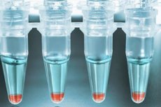
The biological material for the test is a scraping from the posterior-lateral vault of the vagina, cervical canal or urethra. It is taken in a laboratory (gynecological office). For this, the woman needs to prepare in advance. Eubiotics and probiotics should not be taken for 14 days. Sexual intercourse should be avoided approximately 2 days before the test, and tampons should not be used for 24 hours. At least 48 hours should pass after the colposcopy or ultrasound.
How to take Femoflor?
A scraping is taken from the vagina and cervical canal directly during the examination, so the woman does not have to do anything on her own. The only thing that is required is to follow a few simple rules of preliminary preparation. Before the procedure, no hygienic procedures should be performed, and some medications, suppositories, in particular, should not be treated with antiseptics.
Most often, a scraping is taken from the posterior-lateral vaginal vault. In this case, the collection should be done immediately, before the examination. If necessary, excess plaque is removed with a special swab. It is also necessary to maintain the sterility of all the material being examined. Taking the material is an important stage of diagnostics, since if it is violated, you can get a fundamentally incorrect picture of the pathology.
To take a scraping, use a probe, which is then placed in a test tube. The test tube must be labeled and tightly closed with a lid.
Smear for determination of biocenosis of the urogenital tract femoflor
To conduct the study, biological material must be taken. Before starting the collection, it is necessary to refrain from urinating for at least 2 hours before the procedure. The probe is inserted in one movement by approximately 1-1.5 cm, after which it is removed. After the biological material has been collected, the test tube must be labeled. Then the obtained biological material is transported to the laboratory, accompanied by a laboratory technician specializing in transporting samples. Freezing of the sample for a period of no more than 1 month is allowed.
Who to contact?
Technique Femoflor screen
After collecting the biological material, it is delivered to the laboratory, where it is further examined. The purpose of the study is to assess the total bacterial mass, and a quantitative and qualitative assessment of the normal and optional flora is also carried out. Then the percentage of different forms of microorganisms and their relation to the total amount of microflora is assessed.
The principle of the method is based on PCR (polymerase chain reaction) - a molecular genetic method aimed at identifying the DNA strands of the present microflora. This means that it is possible to clearly identify the bacteria, and the result will be 100% reliable.
The advantage of the method is that even a minimal amount of material can be taken for the study and this will be sufficient. This is explained by the fact that the PCR method performs multiple copies of the detected DNA and further identification and analysis of the main properties of the biological material.
The sequence of the procedure can be represented as 4 main stages. First, the DNA strands are unraveled. After that, annealing occurs, during which special primers are attached, onto which nucleic acids are subsequently layered. Then complementary DNA strands are built up. Thus, no matter how much biological material is taken for research, it will be multiplied using PCR, as a result of which it will be possible to fully analyze it.
Femoflor from the cervical canal
Biological material is taken from the cervical canal if there is a suspicion of pathological processes developing in the cervix, or if there are inflammatory processes. If the pathological area is clearly visible in the mirrors, the material is taken from it for examination. After all impurities have been removed, the cervix is treated with a sterile saline solution. The sample is taken using a special probe, which makes circular movements along the entire canal.
How long does it take to do femoflor?
Due to the fact that there is no need for complete cultivation, growth and further identification of microorganisms by biochemical and immunological methods, the speed of the study is significantly increased, and the time spent on conducting a full range of bacteriological studies is reduced. On average, the study is carried out from 1 to 3 working days (unlike standard bacteriological methods, in which the study lasts from 7 to 10 days).
Normal performance
Representatives of the Doderlein group should predominate. Of these, the main ones are lactobacilli, the concentration of which reaches 108-109 CFU/ml. The second place in terms of numbers is occupied by bifidobacteria, the concentration of which fluctuates between 105 and 107 CFU/ml. Propionebacteria are present in a concentration of 104 to 106 CFU/ml.
Presented are eubacteria, clostridia, peptococci, velionella – representatives of obligate flora, as well as representatives of microorganisms of the genus Peptosreptococci (10 4 CFU/ml).
Indigenous flora is represented by aerobic and facultative-anaerobic microorganisms, the concentration of which fluctuates within 10 3 -10 4 CFU/ml. This group includes staphylococci, streptococci, E. coli, enterococci, the number of which fluctuates within 10 3 to 10 4 CFU/ml. The number of Corynebacteria and Klebsiella should not exceed 10 3 CFU/ml.
The device for analysis
The Femoflor kit is required for the procedure. To amplify the detected DNA, a specific set of reagents is required: a DNA matrix, i.e. the area to be amplified, 2 complementary primers, with the help of which the completion will occur. The enzyme thermostable DNA polymerase, which catalyzes the polymerization reaction, is required. Deoxyribonucleophosphate is used as a building material. Magnesium salts and a buffer solution are required for DNA polymerase to work.
The Femoflor reagent kit contains a complex that allows you to calculate the total number of bacteria; a special complex for detecting the composition and number of bacteria that are part of the normal microflora. Additionally, the kits include complexes for determining the composition of facultative microflora. The composition depends on the type of kit (Femochlor 4, 8, 16, 24, etc.).
Raising and lowering of values
In order to decipher the analysis, it is necessary to decipher each block of tests separately, as well as to evaluate the nature of the relationships and the percentage ratio between the different groups.
First, the quality of the biological material is assessed. In the obtained sample, the number of epithelial cells should not exceed 10 4. The total bacterial mass implies determining the volume of all microorganisms in the biocenosis. This has an important diagnostic significance for further assessment of the population ratio. This number should not exceed 10 6. The number of lactobacilli should be maximum - approximately 10 9 CFU/ml. Opportunistic pathogens should not exceed 3-4% of the total bacterial mass. Pathogenic forms can be present in a single form, but this always implies the presence of an infectious process, or a high risk of its development.
Absolute normocenosis
Vaginal microflora is mostly determined by the hormonal background of a woman's body, the nature of which changes and largely depends on the state of the body. This provides selective advantages in the biotope for various forms of microorganisms, which are currently more adapted. The biotope is mainly populated by vaginal biovariants of saccharolytic microorganisms, united under the term "Doderlein". During the development of these microorganisms, a large amount of lactic acid is formed, which prevents the colonization of the biotope by acid-sensitive microorganisms, so the species composition of the biocenosis is quite uniform: the leading positions are occupied by lactic acid lactobacilli, which make up to 97%. The second place among the physiological bacteria of the vagina is occupied by a representative of the genus Bifidobacteria. They have strict anaerobicism, their concentration in the vagina is significantly lower. And finally, the third place is given to representatives of the genus Propionebacteria. Among them, there are strains that have antiviral properties.
Conditional normocenosis
There is also optional microflora, which is represented by opportunistic forms. Their level should not reach 3-4%. The vagina contains up to 20 types of opportunistic pathogens that become active when immunity is reduced.
Types of femoflor
The analysis has several varieties. One or another form is chosen depending on the purpose of the study. Thus, some of them are optimal for studying the urogenital tract when planning pregnancy, operations, which allows identifying potential risks and predicting the further course of the process. Others are used to diagnose venereal infections and monitor the quality of treatment, its effectiveness. They can be used for both men and women.
Others are used to detect a narrow range of infections. For example, femoflor 4 allows you to determine the total concentration of bacteria and identify gardnerella, candida, lactobacilli. The variety of tests varies from femoflor 4 to femoflor 24. The number shows the number of microorganisms that can be detected using these tests. Thus, femoflor 4 allows you to evaluate 4 parameters, while using femoflor 24, you can identify 24 microorganisms.
- Femoflor 4
A highly accurate diagnostic method using the PCR method. Allows you to evaluate 4 main indicators, determine the total biomass, identify representatives of the genus Gardnerella, Candida, Lactobacilli. Based on the results obtained, the laboratory technician draws conclusions about the presence or absence of pathogenic forms of microorganisms. The amount of normal microflora is estimated by the ratio of lactobacilli to TMC - total microbial count.
- Femoflor 8
A method that allows you to study a woman's microbiocenosis and identify 8 main indicators. Allows you to quite effectively identify dysbacteriosis, assess its severity. Usually, this method is sufficient for diagnosing dysbacteriosis and identifying inflammatory processes, assessing the quality of treatment.
- Femoflor 9
It is a method of studying microflora. It is very similar to the femoflor 8 method and has the same indications for implementation. The only difference is that it can be used to detect cytomegalovirus and herpes virus type 2.
- Femoflor 10
The examination is necessarily carried out taking into account the menstrual cycle (in the first half, but not in the first 5 days). It is carried out in case of pronounced symptoms of dysbacteriosis, inflammatory and infectious process. The indication may be both the patient's subjective sensations and objective ones, clearly visible during the examination. It can also be carried out for preventive purposes. It is an extended study of microflora. The results are presented in genome equivalents, the number of which is directly proportional to the cellular biomass of microorganisms.
- Femoflor 12
This is a screening study of the vaginal microbiocenosis, which is carried out using the PCR method. It allows you to quantitatively determine the composition of the microflora. Both obligate and opportunistic microflora are assessed. It is possible to identify some absolutely pathogenic microorganisms, including: Candida, mycoplasma, cytomegalovirus and herpesvirus, as well as Trichomonas, Neisseria, and Chlamydia.
Femoflor 13
This is also a screening test that allows you to evaluate the nature of the microflora and detect pathogenic forms of microorganisms. It is also used to diagnose many hidden infections, such as: ureaplasma, microplasma, chlamydia, and others. The disadvantage of this method is that it only conducts a qualitative assessment of the microbiota, with the exception of the total number of bacteria.
Femoflor 16
This is a study that is most often used to assess the condition of the urogenital tract. The method can also be used for men. With its help, you can not only assess the condition of the microflora, but also identify most venereal infections.
Femoflor 17
Allows to identify 17 different types of microorganisms. Differs from all previous methods in that it allows to fully evaluate both the quantitative and qualitative composition of microflora. Used to evaluate ureaplasma and mycoplasma infections.
Femoflor 18
This is a screening test for urogenital microflora, a modified version of Femoflor 17, which is additionally capable of detecting viral infections.
Femoflor 24
This is the most extensive version of a screening study of the urogenital tract, which allows for the diagnosis of 24 types of microorganisms.
Differences between PCR and Femoflor
Femoflor is a comprehensive study that a doctor can prescribe to a patient if there is a need to identify infections. It is used for preventive purposes, to make a diagnosis and select the appropriate treatment. If there is a need to assess how effective the treatment is, this method is also used. Depending on the type of study, when prescribing a femoflor analysis, the laboratory technician knows what exactly needs to be done and what elements to look for in the sample obtained. It includes a full range of necessary activities, from preparation and collection of biological material to issuance and interpretation of results. One of the most important stages is laboratory diagnostics, during which the PCR method is used to detect the DNA of the microorganism and its subsequent identification.
That is, PCR is one of the methods of laboratory diagnostics, which enables the laboratory technician to detect the DNA of a microorganism and conduct its qualitative and quantitative characteristics. With the help of PCR, it is possible to study any microorganisms, proteins, DNA strands. It has a wide range of possibilities. In the analysis of "femoflor", theoretically, any method could be used, for example, genomic sequencing, RIF, ELISA, and other methods. The result will not change.
Another difference is that a doctor can prescribe a test such as femoflor. But there is no such thing as "prescribe a PCR test", since this is not a test, but a method of conducting a laboratory test, which a sample is subjected to in the laboratory. Depending on the conditions, another method may be chosen, which the laboratory technician considers more rational in a given situation.
For the doctor and the patient, it does not matter what method the study is performed by. The main thing is to get an accurate and correct result. Today, most laboratories use this method, since it has proven itself as reliable, specific, and highly sensitive. The probability of errors is minimal, reliability and accuracy are high.
Florocenosis and femoflor: Which is better?
It is better to use femoflor, since it is an accurate and proven study that allows you to get a complete picture of the urogenital microbiocenosis, identify pathology, and determine its cause. Thus, the doctor will have almost all the necessary data to develop tactics and strategy for further treatment. In addition, there is always the opportunity to repeat the analysis and evaluate the effectiveness of the treatment.