Medical expert of the article

New publications
Amyloidosis of the liver
Last reviewed: 29.06.2025

All iLive content is medically reviewed or fact checked to ensure as much factual accuracy as possible.
We have strict sourcing guidelines and only link to reputable media sites, academic research institutions and, whenever possible, medically peer reviewed studies. Note that the numbers in parentheses ([1], [2], etc.) are clickable links to these studies.
If you feel that any of our content is inaccurate, out-of-date, or otherwise questionable, please select it and press Ctrl + Enter.
Amyloidosis is usually a systemic, general pathology characterized by the accumulation of amyloid (a specific glycoprotein) in tissues and subsequent disruption of normal organ function. Liver amyloidosis is much less common than kidney and spleen [1] but almost always accompanies systemic damage to the body. None of the existing imaging techniques can specifically demonstrate the presence of amyloid. Even when suspected clinically and radiologically, the diagnosis of amyloidosis depends on tissue biopsy to confirm the presence of amyloid deposits. [3] Treatment is complex, comprehensive, and includes immunosuppressive and symptomatic measures. In severe cases, liver transplantation may be required.
Epidemiology
The success of treatment directly depends on the timely diagnosis of the disease, which causes the formation of a protein-polysaccharide complex (amyloid) in various organs and the liver. As practice shows, amyloidosis is difficult to assume or suspect, although it is possible to identify and confirm it. The fact is that in more than 80% of unrecognized cases, the disease is clinically masked by hepatic pathology. The most effective diagnostic method is biopsy.
Liver amyloidosis is a rarer problem when compared to renal amyloidosis. At the same time, all cases of hepatic lesions are accompanied by lesions of other organs. Most often, pathology affects predominantly structural parts of the hepatic triad, which determines the minimum and nonspecificity of symptomatology. Clinical and morphological picture of hepatocellular deficiency and portal hypertension is manifested in diffuse and intralobular type of pathology.
A liver biopsy is justified when hepatomegaly is present without previous hepatic symptoms and in the absence of nephrotic syndrome.
Diffuse liver involvement is seen in about 25% of cases, and in 75% of patients only the portal tracts are affected.
Primary amyloidosis affects the liver in 90% of cases, while secondary amyloidosis affects the liver in only 47% of cases.
Isolated liver involvement is extremely rare. Kidneys (about 93% of cases), spleen (72%), heart (57%), pancreas (36%), adrenal glands (29%), intestines and lungs (21% each) are usually affected synchronously.
Women get the disease almost twice as often as men. The average life expectancy of amyloidosis patients is 52-64 years.
Causes of the hepatic amyloidosis
Amyloidosis proceeds with the formation and accumulation of a complex polysaccharide-protein complex - amyloid - in the liver tissue. The problem of the occurrence of the primary lesion to date is insufficiently studied. As for secondary pathology, its appearance is usually associated with such diseases:
- Chronic infectious processes (tuberculosis, syphilis, actinomycosis);
- Purulent inflammatory processes (microbial endocarditis, osteomyelitis, bronchiectatic disease, etc.);
- Malignant diseases (leukemia, visceral cancer, lymphogranulomatosis).
The reactive form of amyloidosis is found in patients with concomitant atherosclerosis, rheumatologic diseases (Bechterew's disease, rheumatoid arthritis), psoriasis, chronic inflammatory and multisystem processes (including sarcoidosis). The main risk factors: hereditary predisposition, cellular immunity disorders, hyperglobulinemia.
Pathogenesis
There are a number of assumptions regarding the origin of liver amyloidosis. Most specialists adhere to the version of dysproteinosis, immunologic and mutational nature of the disease, as well as local cellular genesis. The version of cellular genesis includes changes in reactions working at the cellular level (formation of fibrillar precursors of amyloid by a complex of macrophages), although amyloid is formed and accumulates outside the cellular structures.
The version of dysproteinosis is based on the fact that amyloid is a product of improper protein metabolism. The basic pathogenetic link of the problem lies in dysproteinemia and hyperfibrinogenemia, which lead to the accumulation of coarse dispersed protein and paraprotein fractions in the plasma.
According to the immunologic version, amyloid formation is caused by antigen-antibody reaction, where tissue decay products or foreign proteins act as antigens. Amyloid accumulation is found mainly in the area of antibody formation and excessive presence of antigens.
The most plausible version scientists consider the mutation theory, which takes into account a variety of mutagenic factors that can lead to abnormalities in protein synthesis.
Amyloid is a complex hypoprotein that consists of globular and fibrillar proteins combined with polysaccharides. Amyloid accumulations affect the intima and adventitia of the vascular network, the stroma of parenchymatous organs, the structure of glands, etc. Amyloid accumulations do not cause functional damage. Small accumulations do not cause functional disorders, but with intense amyloid presence of the liver increases in volume, changes the appearance of the organ, develops a lack of function.
Liver amyloidosis is characterized by the deposition of amyloid fibrils in the space of Dysse, which usually begins in the periportal region, although it is sometimes centrilobular and may also deposit in the hepatic vasculature. [4], [5] In severe cases, amyloid deposition leads to pressure atrophy of hepatocytes, which prevents the passage of bile, resulting in cholestasis, or may block the sinusoids, resulting in portal hypertension. [6], [7], [8]
Symptoms of the hepatic amyloidosis
The clinical picture in liver amyloidosis is diverse, depends on the intensity of amyloid accumulation, on its biochemical features, duration of the pathologic process, the degree of organ damage and violation of their functional state.
At the latent stage of amyloidosis, when amyloid accumulations in the liver can be detected only by microscopic examination, the first signs of the disease are absent. With further development and increasing functional deficit of the organ, the symptomatology progresses.
The liver gradually thickens, enlarges. The method of palpation can be palpated altered, but smooth and painless borders of the organ. Rarely, pathology is accompanied by pain in the subcostal area on the right side, dyspepsia, enlargement of the spleen, yellowing of the skin, mucous membranes and sclerae, hemorrhagic syndrome.
The most characteristic symptoms in liver amyloidosis: [9], [10]
- Amyloid accumulation in the liver causes hepatomegaly in 33-92% of patients;
- Mild jaundice
- Portal hypertension;
- Moderate to severe cholestasis.
Because amyloidosis very rarely affects only one organ, additional symptomatology is usually present:
- When kidney damage develops nephrotic syndrome and arterial hypertension with further renal failure, edema, sometimes renal vein thrombosis, leukocyturia, hematuria, hypoproteinemia, azotemia and so on;
- When the heart is affected, a condition similar to restrictive cardiomyopathy develops (rhythm disturbances, cardiomegaly, increasing cardiac deficit, weakness and dyspnea, edema, less often - fluid accumulation in the abdominal and pleural cavity, pericarditis);
- If the digestive tract is affected, macroglossia, weakness and esophageal peristalsis, nausea and heartburn, constipation or diarrhea, etc. May occur;
- When the pancreas is affected, the symptoms of chronic pancreatitis are present;
- If the musculoskeletal mechanism is involved, symmetrical polyarthritis, carpal tunnel syndrome, myopathies develop, and if the nervous system is affected, polyneuropathies, paralysis, orthostatic low blood pressure, increased sweating, dementia are found.
If the pathologic reaction spreads to the skin, numerous waxy plaques appear on the face, neck, skin folds. The picture of neurodermatitis, red squamous fever, scleroderma is possible.
The combination of multiple amyloid lesions and the variety of symptomatology make the identification of hepatic amyloidosis much more difficult and require a comprehensive and complete diagnosis.
Forms
According to the WHO classification, five types of amyloidosis are distinguished:
- AL (primary);
- AA (secondary);
- ATTR (hereditary and senile systemic);
- Aβ2M (in patients on hemodialysis);
- AIAPP (in patients with insulin-independent diabetes mellitus);
- AB (for Alzheimer's disease);
- AANF (senile atrial amyloidosis).
There is a local amyloidosis of the liver, but more often it is a systemic lesion, in which the pathological process also involves the kidneys, heart, spleen, nervous system, as well as other organs and tissues.
Complications and consequences
Systemic amyloidosis gradually leads to the development of acute pathologic processes that can, in turn, lead to death. Among the most common and life-threatening complications are the following:
- Frequent infectious (bacterial, viral) pathologies, including pneumonias, pyelonephritis, glomerulonephritis;
- Chronic hepatic and renal failure;
- Chronic heart failure (may precede myocardial infarction);
- Hemorrhagic strokes.
Venous thrombosis occurs as a result of accumulation and deposition of proteins on the venous walls. The lumen of the affected vessels narrows, organ failure develops. Over time, against the background of long-term hyperproteinemia, the vessel can completely close off. Any of the complications can lead to an unfavorable outcome - death.
Diagnostics of the hepatic amyloidosis
If liver amyloidosis is suspected, diagnostic measures are carried out after mandatory consultations, both gastroenterologist and therapist, and rheumatologist, cardiologist, dermatologist, neurologist, urologist. It is important to comprehensively evaluate the data of anamnesis and clinical manifestations, to conduct a comprehensive laboratory and instrumental diagnostics.
The tests necessarily include urine and blood examination. In hepatic amyloidosis, a combination of leukocyturia with proteinuria and cylindruria is often found, and hypoproteinemia - with hyperlipidemia, anemia, hyponatremia and hypocalcemia, reduced platelet count. Paraproteins are detected in urine and serum electrophoresis.
Instrumental diagnostics include:
- EKG, Echo;
- Abdominal ultrasound;
- X-rays of the stomach, esophagus;
- Irrigography, barium x-rays;
- Endoscopy.
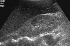
Radiologic findings of hepatic amyloidosis include nonspecific hepatomegaly, increased echogenicity on ultrasound or density on computed tomography (CT), and increased T1 signal intensity on magnetic resonance imaging (MRI). [12] Scintigraphy with Tc-99m-related indicators shows heterogeneous uptake, but it is nonspecific. [13], [14] GC has been shown to increase liver stiffness measured by elastography; [15], [16], [17] but there are few case reports. Magnetic resonance elastography (MRE) is currently the most accurate non-invasive method to detect and stage liver fibrosis, [18], [19] MRE is useful for detecting progression, response to treatment and predicting hepatic decompensation in patients with liver fibrosis. [20]
Amyloidosis of the liver on ultrasound is difficult to determine: an enlargement of the organ is determined, with the most specific hepatomegaly exceeding 15 cm. Under the control of ultrasound, a biopsy is performed, which becomes a determining indicator for diagnosis. Using a special needle, a small amount of liver tissue is taken, then it is stained with a special dye and examined under a microscope, which allows you to directly see amyloid deposits.
A definitive diagnosis is made only after the detection of amyloid fibrils in the tissue of the liver and other organs. The genetically determined type of amyloidosis is determined by careful genetic-medical examination of the pedigree.
Differential diagnosis
Amyloidosis should be suspected in all patients with a combination of renal proteinuria, restrictive cardiomyopathy, autonomic or peripheral neuropathy, and hepatomyelia, even in the absence of monoclonal paraprotein. Verifying the type of amyloidosis is very important because the treatment of lesions of different etiologies is very different.
Histological diagnosis involves staining with Congo red followed by microscopic examination in polarizing light. It is advisable to biopsy several tissue samples at once. If the result of staining becomes positive, immunohistochemical analysis is performed using monoclonal antibodies to precursor proteins to identify the type of amyloid.
DNA analysis is performed to differentiate between primary amyloidosis and different variations of genetically determined amyloidosis. Amyloid fibrils can be isolated from biopsy specimens and sequestered into individual amino acids.
Additional studies to determine plasma cell dyscrasia:
- Electrophoresis of serum proteins of blood and urine;
- Immunoassay for free light chains;
- Immunofixation (immunoblotting) of serum proteins;
- Bone marrow aspiration and trepanobiopsy.
Diagnosis of liver amyloidosis is a lengthy and labor-intensive process, which requires increased attention of specialists and quality equipment of clinics and laboratories.
Who to contact?
Treatment of the hepatic amyloidosis
Treatment measures are aimed at reducing the concentration of pre-existing amyloid proteins in the blood (eliminating the cause of amyloidosis) and supporting adequate liver function.
Secondary amyloidosis requires blocking the inflammatory process (in chronic infectious and autoimmune pathologies). In autoimmune diseases, the use of cytostatics is recommended. To eliminate chronic infectious processes, the area of inflammation is often surgically removed. Often this approach can stop further progression of amyloidosis and improve liver function.
Primary amyloidosis requires the use of chemopreventive drugs and sometimes bone marrow transplantation.
Current guidelines recommend the combination of cyclophosphamide, bortezomib, dexamethasone (CyBorD), and daratumumab as first-line therapy in patients newly diagnosed with AL.
Bortezomib is a proteasome inhibitor. Proteasomes are involved in reducing proteotoxicity and regulating proteins that control cellular progression and apoptosis. Plasma cells that generate amyloid are particularly sensitive to proteasome inhibition because they rely on the proteasome to reduce the toxic effects of light chains and prevent apoptosis.
Daratumumumab is a monoclonal antibody (mAb) that binds to CD38, a transmembrane glycoprotein expressed on the surface of plasma cells, inducing apoptosis. It is the only drug specifically approved for the treatment of AL amyloidosis when used with CyBorD. The efficacy of CyBorD-daratumumumab is very high, with 78% of patients achieving a significant hematologic response (defined as a complete response or very good partial response). Median survival in the small group of patients receiving CyBorD ( n = 15) was 655 days compared to 178 days for patients receiving other melphalan-dexamethasone-based treatment ( n = 10). 4
However, these therapies have numerous side effects, including cardiotoxicity, leading to the need for dose reduction or suspension of treatment, and the use of other less effective but more tolerable therapeutic strategies.
Isatuximab, a monoclonal antibody against CD38 similar to daratumumab, is being studied for the treatment of plasma cell dyscrasia underlying AL.
Three monoclonal antibodies birtamimab, CAEL-101 and AT-03 are currently being studied for the removal of amyloid fibrils from diseased organs. The results of these studies will be able to offer direct evidence for the hypothesis that by removing light chain deposition fibrils from organs there is an improvement in organ function. [21]
In order to support liver function, drugs based on urso-deoxycholic acid are prescribed (example - Ursosan). Urso-deoxycholic acid helps stabilize cell membranes, reduces the adverse effect of toxic fatty acids in bile stasis provoked by amyloid deposits, and helps restore normal bile outflow.
In addition, symptomatic therapy and support for the functioning of other vital structures such as the nervous system, heart, kidneys, etc. Supportive therapy for patients with hepatic amyloidosis includes various clinical aspects, including treatment of heart failure, arrhythmias, conduction disorders, thromboembolism and the concomitant presence of aortic stenosis.
Other treatments depend on the type of amyloidosis and which parts of the body are affected. Treatments may include: [22]
- Medicines that relieve symptoms, such as pain relievers, nausea medicines, or medicines that reduce swelling (diuretics);
- Medications to reduce amyloid;
- Kidney dialysis;
- Liver transplant.
The liver produces 95% of TTR (transthyretin, a protein involved in thyroxine (T4) transport and retinol-binding protein. Transthyretin is mainly synthesized in the liver and is rich in beta strands that tend to aggregate into insoluble amyloid fibrils) measured in serum. Therefore, liver transplantation has historically (since 1990) been suggested as first-line therapy to eliminate the major source of amyloidogenic TTR in patients with the familial form (ATTRv), whereas it is not indicated in the ATTR-wt form. Liver transplantation of young patients in the early stages of the disease is associated with a high 20-year survival rate. Liver transplantation appears to be more effective in some mutations and less effective in others, such as V122I (associated with cardiomyopathy). Combined liver and heart transplantation is also possible in young ATTRv patients with cardiomyopathy, and literature data on a small group of patients suggest that this combination has a better prognosis than heart transplantation alone.
Patients with hepatic amyloidosis are contraindicated taking cardiac glycosides and calcium antagonists such as Diltiazem or Verapamil, which can accumulate in amyloid. ACE inhibitors and beta-adrenoblockers are used with caution.
In orthostatic hypotension, mineralocorticoids or glucocorticosteroids are prescribed, taking into account that they can cause decompensation of heart failure. The alpha-adrenomimetic midodrine (Gutron) is also used with caution.
Anticonvulsants and antidepressants are appropriate in neuropathies.
In some cases of liver amyloidosis, doctors have to consider transplantation of the organ.
Prevention
Due to the lack of information about the pathogenesis of liver amyloidosis, specialists can not develop a specific prevention of the disease. Therefore, the main efforts are reduced to the timely detection and treatment of any chronic pathologies that can provoke the development of the disorder. If there are cases of amyloidosis of any localization in the family, it is recommended to systematically visit doctors for dispensary examinations.
In general, preventive measures are reduced to the timely elimination of infectious diseases, especially those that tend to transform into a chronic process. It is about preventing the development of tuberculosis, pulmonary infections, etc. It is important to timely detection and adequate treatment of streptococcal infections, which can become the cause of chronic forms of autoimmune inflammatory processes. We are talking about scarlatina, streptococcal tonsillitis, etc.
If the patient already has an autoimmune disease, then he should systematically consult with a doctor, observe the activity of the pathology, apply the necessary medications as prescribed by the doctor, adjust dosages according to indications.
Forecast
The prognosis for patients with hepatic amyloidosis is unfavorable. The disease increases slowly but continuously, which eventually causes dysfunction of the affected organs and lethal outcome - in particular, due to organ failure.
Patients with systemic pathology mainly die as a result of the development of chronic renal failure, although in some cases hemodialysis or continuous ambulatory peritoneal dialysis improves the prognosis of such patients. The survival rate of patients on hemodialysis, regardless of its type, can be compared with that of people with other systemic pathologies and diabetes mellitus.
The main cause of death during hemodialysis is the development of complications from the cardiovascular system.
Liver transplantation has long been considered one of the main methods of treatment of the disease, and the most optimistic survival rates are observed in patients whose age does not exceed 50 years (provided that the pathological process is short-lived and the body mass index is normal). Patients with liver amyloidosis combined with peripheral neuropathy have somewhat worse prognosis.