Medical expert of the article

New publications
Respiratory distress syndrome in adults
Last reviewed: 05.07.2025

All iLive content is medically reviewed or fact checked to ensure as much factual accuracy as possible.
We have strict sourcing guidelines and only link to reputable media sites, academic research institutions and, whenever possible, medically peer reviewed studies. Note that the numbers in parentheses ([1], [2], etc.) are clickable links to these studies.
If you feel that any of our content is inaccurate, out-of-date, or otherwise questionable, please select it and press Ctrl + Enter.
Adult respiratory distress syndrome (ARDS) is an acute respiratory failure that occurs with acute lung injury of various etiologies and is characterized by non-cardiogenic pulmonary edema, respiratory failure, and hypoxia.
The syndrome was described by Esbach in 1967 and named by analogy with neonatal distress syndrome, which is caused by congenital surfactant deficiency. In adult respiratory distress syndrome, surfactant deficiency is secondary. Synonyms for adult respiratory distress syndrome are often used in the literature: shock lung, non-cardiogenic pulmonary edema.
According to Marini (1993), 150,000 cases of adult respiratory distress syndrome are registered annually in the USA, which is 0.6 per 1000 population.
Cause of Adult Respiratory Distress Syndrome
The most common causes of adult respiratory distress syndrome are:
- pneumonia (bacterial, viral, fungal and other etiologies);
- sepsis;
- shock (septic, anaphylactic, etc.), long-lasting and severe;
- disseminated intravascular coagulation syndrome (acute and subacute course);
- aspiration of vomit, water (in case of drowning);
- chest trauma and compartment syndrome;
- inhalation of irritating and toxic substances: chlorine, nitrogen oxides, phosgene, ammonia, pure oxygen (oxygen intoxication);
- pulmonary embolism (fat, air, amniotic fluid);
- massive blood transfusions, which cause multiple microthromboemboli in the pulmonary vascular bed. This is due to the fact that in preserved blood up to 30% of erythrocytes are in the form of microaggregates up to 40 μm in diameter, and the lungs, being a kind of filter, retain these microaggregates and the pulmonary capillaries become clogged. In addition, serotonin is released from erythrocytes, causing spasm of the pulmonary arterioles and capillaries;
- venous fluid overload (colloidal and saline solutions, plasma, plasma substitutes, fat emulsions);
- use of artificial blood circulation apparatus (postperfusion respiratory distress syndrome in adults);
- severe metabolic disorders (diabetic ketoacidosis, uremia);
- acute hemorrhagic pancreatic necrosis. In the development of adult respiratory distress syndrome in acute pancreatitis, enzyme intoxication is of great importance, causing a disruption in surfactant synthesis. A particularly large role is given to the enzyme lecithinase A, which intensively destroys surfactant, which leads to the development of alveolar atelectasis, obliterating alveolitis, and predisposes to the development of pneumonia;
- autoimmune diseases - systemic lupus erythematosus, Goodpasture's syndrome, etc.;
- prolonged stay at high altitude.
Pathogenesis of adult respiratory distress syndrome
Under the influence of etiological factors, a large number of activated leukocytes and thrombocytes accumulate in the pulmonary capillaries and interstitial tissue of the lungs. It is assumed that they release a large number of biologically active substances (proteinases, prostaglandins, toxic oxygen radicals, leukotrienes, etc.), which damage the alveolar epithelium and vascular endothelium, change the tone of the bronchial muscles, vascular reactivity, and stimulate the development of fibrosis.
Under the influence of the above-mentioned biological substances, the endothelium of the pulmonary capillaries and alveolar epithelium is damaged, vascular permeability increases sharply, pulmonary capillaries spasm and the pressure in them increases, there is a pronounced exudation of plasma and erythrocytes into the alveoli and interstitial tissue of the lungs, pulmonary edema and atelectasis develop. The development of atelectasis is also facilitated by a secondary decrease in surfactant activity.
As a result of the named processes, the main pathophysiological mechanisms develop: hypoventilation of the alveoli, shunting of venous blood into the arterial bed, disruption of the correspondence between ventilation and perfusion, disruption of the diffusion of oxygen and carbon dioxide.
 [ 6 ], [ 7 ], [ 8 ], [ 9 ], [ 10 ], [ 11 ], [ 12 ]
Pathomorphology of adult respiratory distress syndrome
Adult respiratory distress syndrome develops over a period of time from several hours to 3 days from the onset of exposure to the etiologic factor. There are three pathomorphological phases of adult respiratory distress syndrome: acute, subacute and chronic.
The acute phase of adult respiratory distress syndrome lasts 2-5 days and is characterized by the development of inherstitial and then alveolar pulmonary edema. The edema fluid contains protein, erythrocytes, and leukocytes. Along with edema, damage to the pulmonary capillaries and severe damage to the alveolar epithelium of types I and II are detected. Damage to type II alveolocytes leads to a disruption in surfactant synthesis, resulting in the development of microatelectasis. With a favorable course of adult respiratory distress syndrome, acute symptoms subside after a few days, and the edema fluid is absorbed. However, such a favorable course of adult respiratory distress syndrome is not always observed. In some patients, adult respiratory distress syndrome passes into a subacute and chronic phase.
The subacute phase is characterized by interstitial and bronchoalveolar inflammation.
The chronic phase of adult respiratory distress syndrome is the phase of development of fibrosing alveolitis. Connective tissue grows in the alveolar-capillary basement membrane, the membrane thickens sharply and flattens. There is pronounced proliferation of fibroblasts and increased synthesis of collagen (its amount increases 2-3 times). Pronounced interstitial fibrosis can form in 2-3 weeks. In the chronic phase, changes in the vascular bed of the lungs are also observed - desolation of vessels, development of microthrombosis. Ultimately, chronic pulmonary hypertension and chronic respiratory failure develop.
Symptoms of Adult Respiratory Distress Syndrome
In the clinical picture of adult respiratory distress syndrome, it is customary to distinguish 4 periods. Period - latent or period of influence of the etiologic factor. It lasts about 24 hours after the influence of the etiologic factor. During this period, pathogenetic and pathophysiological changes occur, but they do not have any clinical or radiological manifestations. However, tachypnea is often observed (the number of breaths is more than 20 per minute).
II period - initial changes, develops in 1-2 days from the onset of the action of the etiologic factor. The main clinical symptoms of this period are moderate dyspnea, tachycardia. Auscultation of the lungs can reveal harsh vesicular breathing and scattered dry wheezing.
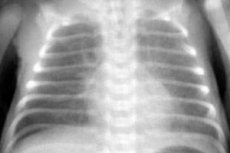
Chest X-rays show an increase in vascular pattern, primarily in the peripheral areas. These changes indicate the onset of interstitial pulmonary edema.
A blood gas analysis either shows no deviations from the norm or reveals a moderate decrease in PaO2.
Period III - a developed period or a period of pronounced clinical manifestations, characterized by pronounced symptoms of acute respiratory failure. Pronounced dyspnea appears, accessory muscles participate in breathing, the flaring of the wings of the nose and retraction of the intercostal spaces are clearly visible, pronounced diffuse cyanosis is observed. During auscultation of the heart, tachycardia and muffled heart sounds are noticeable, arterial pressure decreases significantly.
Percussion of the lungs reveals dullness of the percussion sound, more in the posterior lower sections, auscultation reveals harsh breathing, dry wheezing may be heard. The appearance of moist wheezing and crepitation indicates the appearance of fluid in the alveoli (alveolar pulmonary edema of varying severity).
The chest radiograph shows pronounced interstitial pulmonary edema, as well as bilateral infiltrative shadows of irregular cloud-like shape, merging with the roots of the lungs and with each other. Very often, focal shadows appear in the marginal sections of the middle and lower lobes against the background of an enhanced vascular pattern.
A characteristic feature of this period is a significant drop in PaO2 (less than 50 mm Hg, despite oxygen inhalation).
Period IV is terminal and is characterized by pronounced progression of respiratory failure, development of severe arterial hypoxemia and hypercapnia, metabolic acidosis, and formation of acute pulmonary heart disease due to increasing pulmonary hypertension.
The main clinical symptoms of this period are:
- severe shortness of breath and cyanosis;
- profuse sweating;
- tachycardia, muffled heart sounds, often various arrhythmias;
- a sharp drop in blood pressure, even to the point of collapse;
- cough with the production of foamy pink sputum;
- a large number of moist rales of varying caliber in the lungs, abundant crepitation (signs of alveolar pulmonary edema);
- development of signs of increasing pulmonary hypertension and acute pulmonary heart syndrome (splitting and accentuation of the second tone in the pulmonary artery; ECG signs - high pointed P waves in leads II, III, avF, V1-2, pronounced deviation of the electrical axis of the heart to the right; radiological signs of increased pressure in the pulmonary artery, bulging of its cone);
- development of multiple organ failure (impaired renal function, which is manifested by oliguria, proteinuria, cylindruria, microhematuria, increased blood urea and creatinine levels; impaired liver function in the form of mild jaundice, significant increase in blood alanine aminotransferase, fructose-1-phosphate aldolase, lactate dehydrogenase; impaired brain function in the form of lethargy, headaches, dizziness, possible clinical signs of cerebrovascular accident).
Blood gas analysis reveals profound arterial hypoxemia, hypercapnia, and acid-base balance analysis reveals metabolic acidosis.
Where does it hurt?
What's bothering you?
Diagnosis of adult respiratory distress syndrome
In 1990, Fisher and Foex proposed the following diagnostic criteria for adult respiratory distress syndrome:
- respiratory failure (severe shortness of breath);
- increased work of breathing, increasing rigidity of the chest;
- clinical picture of increasing pulmonary edema;
- typical radiological picture (increased pulmonary markings, interstitial pulmonary edema);
- arterial hypoxemia (usually PaO2 less than 50 mmHg) and hypercapnia;
- hypertension in the pulmonary circulation (pressure in the pulmonary artery is more than 30/15 mm Hg);
- normal pulmonary artery wedge pressure (<15 mm Hg). Determination of this criterion is important for differentiating adult respiratory distress syndrome from cardiogenic pulmonary edema, which is characterized by an increase in pulmonary artery wedge pressure;
- Arterial blood pH is less than 7.3.
Screening program for adult respiratory distress syndrome
- General blood and urine analysis.
- ECG.
- X-ray of the lungs.
- Study of acid-base balance.
- Blood gas analysis: determination of PaO2, PaCO2.
What do need to examine?
How to examine?
What tests are needed?