
Trigeminal nerve
Last reviewed: 20.11.2021

All iLive content is medically reviewed or fact checked to ensure as much factual accuracy as possible.
We have strict sourcing guidelines and only link to reputable media sites, academic research institutions and, whenever possible, medically peer reviewed studies. Note that the numbers in parentheses ([1], [2], etc.) are clickable links to these studies.
If you feel that any of our content is inaccurate, out-of-date, or otherwise questionable, please select it and press Ctrl + Enter.
The trigeminal nerve (n. Trigiinus), being a mixed nerve, innervates the skin of the face, the mucous membrane of the nose and its sinuses, the oral cavity, the front 1/3 of the tongue, teeth, conjunctiva eyes, masticatory muscles, muscles of the oral cavity floor (mandibular, sublingual, chin sublingual, anterior abdomen of the digastric muscle), a muscle that strains the eardrum, and a muscle that strains the palatal curtain. The triple nerve has a motor nucleus and three sensitive nuclei (median brain, bridge and spinal cord). From the brain, the trigeminal nerve comes out with two roots - motor and sensory. The sensory spine is much thicker (5-6 mm) than the motor spine (1 mm). Both roots come out of the brain in the area of the bridge to the middle cerebellum pedicle. The sensory radix (radix sensoria) is formed by the central processes of pseudo-unipolar cells, whose bodies are in the trigeminal node. The triple node (ganglion trigeminale; semilunar, gasser node) is located in the trigeminal depression on the anterior surface of the temporal bone pyramid, in the cleft of the hard shell of the brain (in the trigeminal cavity). The node has a semilunar form, its length is 1.4-1.8 cm, the width of the node is 3 times less than the length. The sensory spine is directed to the sensitive nuclei of this nerve. Axons of neurons of sensitive trigeminal nerve nuclei located in the brainstem pass to the other side (form a cross) and are directed to the nerve cells of the thalamus. Peripheral processes of neurons go in the trigeminal nerve and end with receptors in the skin and mucous membranes of the head. The motor spine (radix motoria) of the trigeminal nerve lies below the trigeminal node (not included in it) and participates in the formation of the third branch of the trigeminal nerve.
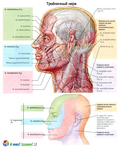
Three large branches leave the trigeminal nerve:
- the optic nerve;
- the maxillary nerve;
- the mandibular nerve.
The ocular and maxillary nerves contain only sensitive fibers, the mandibular nerve is sensitive and motor.
The optic nerve (n. Ophtalmicus) - the first branch of the trigeminal nerve, passes in the thickness of the lateral wall of the cavernous sinus. Together with the oculomotor, block and discharge nerves is directed to the upper orbital fissure. Before entering the eye socket at the level of the Turkish saddle, the eye nerve receives connective branches from the periarterial sympathetic plexus of the internal carotid artery. Here the eye nerve gives the tentorial (shell) branch (r. Tentorii [meningeus]). This branch goes back and ramifies in the hint of the cerebellum, in the walls of the direct and transverse sinuses of the hard shell of the brain. At the entrance to the upper glandule, the optic nerve is medial to the nerve block, higher and lateral to the oculomotor and lateral to the outgoing nerve. Entering the eye socket, the eye nerve is divided into the frontal, nosoresnychny and tear nerves.
The frontal nerve (n. Frontalis) is the longest branch of the optic nerve, it passes under the upper wall of the orbit. On the upper surface of the muscle, lifting the eyelid, the frontal nerve is divided into the supraorbital and suprapubic nerves. The supraorbital nerve (n. Supraorbitalis) exits through the supraorbital notch from the orbit and ends in the forehead skin. The supraclavicular nerve (n. Supratrochlearis) rises above the block of the upper oblique muscle and branches into the skin of the nose, the lower forehead and in the medial angle of the eye, in the skin and conjunctiva of the upper eyelid.
The nosocomial nerve (n. Nasociliaris) passes in the orbit above the optic nerve, between it and the upper rectus muscle of the eye, and then - between the oblique and medial rectus muscles of the eye. Here the nosorozhnichny nerve is divided into its final branches, directed to the conjunctiva of the eye, the skin of the upper eyelid and the mucous membrane of the nasal cavity. In the course of the nosorozhnichny nerve gives a number of branches:
- connecting branch (with ciliary knot) [r. Commiinicans (cum gangliociliari)] - a long spine to the ciliary node. This spine departs from the initial part of the nososnichnichnogo nerve, crosses the obliquely and from above the optic nerve, goes to the ciliary node;
- long ciliary nerves (nn. Ciliares longi) in the form of 2-3 branches pass along the upper surface of the nerve to the back of the eyeball;
- the posterior latticed nerve (n. Ethmoidalis posterior) through the eponymous hole in the medial wall of the orbit penetrates into the thickness of the mucosa of the posterior cells of the latticed bone and the sphenoid sinus;
- the front latticed nerve (n. Ethmoidalis anterior) through the eponymous hole in the medial wall of the orbit penetrates into the cavity of the skull, gives the branch to the hard shell of the brain (in the region of the anterior cranial fossa). Passing forward along the upper surface of the perforated plate, the nerve penetrates through one of its anterior holes into the nasal cavity and branches into the mucous membrane of the nose, the frontal sinus and the skin of the tip of the nose;
- the subfamil nerve (n. Infratrochlearis) runs along the medial wall of the orbit under the upper oblique muscle of the eye to the lacrimal sac, lacrimal flesh, skin of the upper eyelid and to the back of the nose.
The lacrimal nerve (n. Lacrimalis) first passes between the lateral and upper rectus muscles of the eye, then lies near the upper-lateral corner of the orbit. Gives branches to the lacrimal gland, the conjunctiva of the upper eyelid and the skin in the region of the outer corner of the eye. A connecting branch from the zygomatic nerve - the branch of the maxillary nerve - approaches the lacrimal nerve [r. Communicans (cum n. Zygomatici)], which carries secretory parasympathetic fibers for the lacrimal gland.
The maxillary nerve (n. Maxillaris) enters the orbit through the lower orbital gap, lies in the infraorbital furrow, which passes into the infraorbital canal. At the level of the infraorbital furrow and canal, the upper alveolar nerves (nn alveolares superiores), as well as the anterior, middle and posterior alveolar branches (r. Alveolares anteriores, medius and posteriores) extend from the infraorbital nerve . They form the upper dental plexus (plexus dentalis superior), located in the maxillary bone and in the mucosa of the maxillary sinus. The upper dental branches (r. Dentales superiores) to the teeth and the upper gingival branches (r. Gingivales superiores) exit from the plexus to the gums of the upper jaw. The inner nasal branches (rr.nasales interni) from the maxillary nerve also extend to the mucosa of the anterior parts of the nasal cavity.
The infraorbital nerve (n. Infraorbitalis) leaving the infraorbital foramen favors the divergent lower branches of the eyelid (rr. Palpebrales inferiores), the outer nasal branches (rr. Nasales externi), the upper labial branches (r. Labiales superiores, "small crow's foot") . The outer nasal branches in the number of two or three pass through the nasal muscle into the skin of the wing of the nose. Upper labial branches in the number of three to four are directed downward to the mucosa of the upper lip.
The stylus nerve (n. Zygomaticus) departs from the maxillary nerve in the pterygoid-palatine fossa, is directed into the orbit via the upper orbital fissure. In the orbit gives a parasympathetic branch (from the winged nodulus) to the lacrimal nerve, intended for the secretory innervation of the lacrimal gland. In the orbit, the zygomatic nerve passes near its lateral wall, enters the cheek-eyed orifice, where it divides into the cheek-and-cheek-shaped branches. The skeleton branch (r. Zygomaticotiporalis) passes through the cheek-shaped aperture from the zygomatic bone and divides into 2 branches innervating the skin of the anterior part of the temporal region and the lateral forehead.
The skolulitic branch (r. Zygomaticofacialis) usually leaves with two or three stems through the same hole on the face and innervates the skin of the upper part of the cheek and the lateral part of the lower eyelid.
In the pterygo-palatine fossa, the maxillary nerve gives to the winged nodule two or three thin nodal branches (rg. Ganglionares, s. Ganglionici) containing sensitive nerve fibers. A smaller part of the nodular fibers enters directly into the ves- ponent node. A greater number of these fibers go near the lateral surface of the node and pass into its branches.
The pterygopalon (ganglion pterygopalatinum) refers to the parasympathetic part of the autonomic nervous system. It is located in the pterygo-palatine fossa, medially and downward from the maxillary nerve. To the node are suitable (in addition to sensitive, transit branches) preganglionic parasympathetic fibers. They enter the pterygoid node in the form of a large stony nerve (from the facial nerve) and terminate on the neurons that make up the node. Axons of neurons of the node in the form of postganglionic parasympathetic fibers leave the node as part of its branches. The postganglionic sympathetic fibers from the pterygoid nerve are also suitable for the winged nodule. These fibers pass through the vortex node in transit and are part of the branches of this node [cf. "Autonomic (autonomic) nervous system"].
The following branches branch out from the vascular nodule:
- the medial and lateral superior posterior nasal branches penetrate the wedge-palatine foramen in the nasal cavity where the mucosa is innervated. The nasonephalic nerve (n. Nasopalatine) departs from the upper medial branches . It innervates the mucous membrane of the septum of the nose, and after exiting through the incisive canal into the oral cavity - the mucous membrane of the anterior part of the hard palate. Lateral and medial superior posterior nasal branches also extend to the pharyngeal arch, the walls of the khohans and the sinus of the sphenoid bone;
- the large palatine nerve (n. Palatinus major) penetrates through the large palatine opening to the lower surface of the hard palate, innervates the mucous membrane of the gums, the hard palate, including the palatine glands. The nerve also gives the posterior nasal branches (rr. Nasales posteriores inferiores) to the mucosa in the region of the inferior nasal cavity, the middle and lower nasal passages, and also the maxillary sinus;
- small palatine nerves (nn. Palatini minores) through small palatine orifices go to the mucosa of the soft palate and to the palatine tonsil.
The mandibular nerve (n. Mandibularis) - the third, the largest branch of the trigeminal nerve, contains both motor and sensitive fibers. From the cranial cavity the mandibular nerve leaves through the oval aperture and is immediately divided into motor and sensitive branches.
Motor branches of the mandibular nerve:
- masticatory nerve (n. Massetericus);
- deep temporal nerves (nn temporales profundi);
- lateral and medial pterygoid nerves (n., pterygoidei lateralis et medialis). These nerves are sent to the masticatory muscles.
The motor nerve also includes the nerve of the muscle that strains the eardrum (n. Musculi tensoris tympani), and the nerve of the muscle that strains the palatal curtain (n. Musculi tensoris veli palatini).
Sensitive branches of the trigeminal nerve:
- The meningeal branch (r. Meningeus), or the spinous nerve, leaves slightly below the oval aperture, enters through the spinous aperture into the cranial cavity together with the middle meningeal artery and divides into the anterior and posterior branches. The anterior branch innervates the hard shell of the brain. The posterior branch leaves through the stony-scaly slit, innervates the mucous membrane of the cells of the mastoid process of the temporal bone;
- the buccal nerve (n. Buccalis) goes between the lateral and medial pterygoids, perforates the buccal muscle, branched out in the mucous membrane of the cheek, gives branches to the skin in the corner of the mouth;
- the anterior-temporal nerve (n. Auriculotiporalis) with two roots covers the middle meningeal artery. Then, in the form of a single trunk, the nerve goes up, passes through the parotid salivary gland and gives off a series of branches:
- articular branches (r. Articulares) are directed to the capsule of the temporomandibular joint;
- Parotid branches (rr. Parotidei) go to the parotid salivary gland. These branches contain postganglionic parasympathetic (secretory) fibers to the parotid gland;
- the anterior ears (nn. Auriculares anteriores) go to the anterior part of the auricle;
- the nerves of the external auditory canal (nn. Meatus acustici externi) innervate the walls of the external auditory canal at the junction of the cartilaginous and bony parts of it and the tympanic membrane;
- the branches of the tympanic membrane (rr. Mebranae tympani) go to the eardrum;
- the superficial temporal branches (r. Temporales superficiales) go to the skin of the temporal region.
Under the oval aperture on the medial side of the temporomandibular joint is a vegetative earplant (ganglion oticum) of oval form, 3-4 mm long. Preganglionic parasympathetic fibers to the ear node fit in the small stony nerve (from the facial nerve);
- the lingual nerve (n. Lingualis) goes between the lateral and medial pterygoids, then the nerve turns abruptly, passes along the inner surface of the body of the lower jaw between the submandibular salivary gland and the sublingual-lingual muscle upward. Numerous sensitive branches of the lingual nerve end in the mucosa of the anterior Vl of the tongue and in the hyoid area.
The nodal branches also extend from the lingual nerve to the submandibular and sublingual parasympathetic nodes [cf. "Parasympathetic part of the autonomic (autonomic) nervous system"]. These nodes are suitable fibers that join the lingual nerve in the drum string - one of the branches of the facial nerve. The drum string approaches the lingual nerve at an acute angle in its initial part (between the medial and lateral pterygoid muscles). It carries taste fibers that innervate the mucous membrane of the anterior 2/3 of the tongue;
- the lower alveolar nerve (n. Alveolaris inferior) contains the sensory and motor fibers and is the largest branch of the mandibular nerve. This nerve first passes between the medial and lateral pterygoid muscles, then enters the mandibular canal through its inlet on the inner surface of the lower jaw. At the place of entry into the canal from the lower alveolar nerve, the motor branches branch out to the maxillofacial and the chin-hyoid muscles, to the anterior abdomen of the digastric muscle - the jaw-hyoidic branch (r. Mylohyoideus). In the mandibular canal, the lower alveolar nerve (passing along with the same artery and vein) gives off the branches forming the lower dental plexus (plexus dentalis inferior). From the plexus to the teeth of the lower jaw, the lower dental branches (r. Dentales inferiores), and to the gums - the lower gingival inferiores (g.
- after exiting through the chin aperture the lower alveolar nerve passes into the chin nerve (n. Mentalis), which ends in the skin of the chin and lower lip. He gives to them the chin branches (rr. Mentales), the lower labial branches (r. Labiales inferiores), and also the branches to the gums (r. Gingivales).
Where does it hurt?
What do need to examine?
How to examine?