Arthrocenthesis
Last reviewed: 23.04.2024

All iLive content is medically reviewed or fact checked to ensure as much factual accuracy as possible.
We have strict sourcing guidelines and only link to reputable media sites, academic research institutions and, whenever possible, medically peer reviewed studies. Note that the numbers in parentheses ([1], [2], etc.) are clickable links to these studies.
If you feel that any of our content is inaccurate, out-of-date, or otherwise questionable, please select it and press Ctrl + Enter.

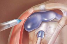
Arthrocentesis is the procedure of needle puncture. With properly performed arthrocentesis and the presence of effusion in the joint, it is possible to obtain the latter for examination. The study of synovial fluid is the most accurate method of determining the cause of effusion and is indicated in all cases of significant amounts of effusion in one or more joints, the cause of which is unclear.
Contraindications
The presence of infection and other skin rashes at the site of the proposed puncture is a contraindication to the procedure.
Method of conducting
Arthrocentesis is performed in aseptic conditions. Before it is carried out, it is necessary to prepare a container for placing the sample. Local anesthesia is performed with lidocaine or a spray of difluoroethane. To avoid damage to the nerves, arteries and veins, which are usually located in the area of the flexor surfaces of the joints, many joints are punctured on the side of the opposite (extensor) surface. For the puncture of most joints, a needle with a diameter of 0.9 mm is used, through which the maximum possible amount of liquid is removed. For the correct procedure, there are a number of anatomical landmarks.
Wrist-phalanx, metatarsophalangeal and interphalangeal joints are identified identically: use a needle 0.8 or 0.7 mm in diameter; Puncture is performed from the back surface on either side of the tendon.
What do need to examine?


 [
[