Seborrheic keratosis: causes, symptoms, diagnosis, treatment
Last reviewed: 23.04.2024

All iLive content is medically reviewed or fact checked to ensure as much factual accuracy as possible.
We have strict sourcing guidelines and only link to reputable media sites, academic research institutions and, whenever possible, medically peer reviewed studies. Note that the numbers in parentheses ([1], [2], etc.) are clickable links to these studies.
If you feel that any of our content is inaccurate, out-of-date, or otherwise questionable, please select it and press Ctrl + Enter.

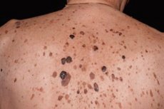
Seborrheic keratosis (syn: seborrheic wart, senile wart, basal cell papilloma, seborrheic nevus Unna, seborrheic keratopapilloma) - benign tumor. Quite a frequent disease that occurs mainly in the second half of life, less often - at a younger age. Localized on the face, trunk. It is a sharply limited hyperpigmented spot with a smooth or slightly scaly surface with a diameter of up to several centimeters, or a plaque or nodular formation with a warty surface and a varying degree of pigmentation, covered with dry horny masses. Can be single, more often plural.
Pathomorphology. Seborrheic keratosis mostly has an exophytic papillomatous type of growth, less often with the spreading into the dermis of massive layers of epithelial cells of various configurations. Histologically distinguish "irritated" (hyperkeratotic), adenoid or reticular, flat (acanthotic) types of seborrheic keratosis. Often, in the same hearth, all types of symptoms can be combined.
Hyperkeratonic type is characterized by acanthosis, hyperkeratosis, papillomatosis. The horny layer in places invaginates into the epidermis, resulting in the formation of cystic cavities filled with horny masses (pseudo-cysts). Acanthotic strands consist mainly of spiny cells, but in places there are accumulations of basaloid cells
The flat (acanthotic) type differs by a sharp thickening of the epidermis with relatively moderately expressed hyperkeratosis and papillomatosis. There is a large number of pseudorandom cysts with a predominance of basaloid-type cells along the periphery.
In the adenoid type, there is a proliferation of numerous narrow branching strands consisting of 1-2 rows of basaloid cells in the upper parts of the dermis. The horny brush is sometimes of considerable size, so you can talk about the alenoid-cystic variant.
In the "irritated" type of seborrhoeic keratosis, a significant inflammatory infiltrate in the dermis is revealed with exocytosis of the cellular elements of the infiltrate into the structures of the neoplasm, which is accompanied by flat-epithelial differentiation and the formation of numerous foci of keratinization of round shape, denoted in the English literature as eddies. The histological pattern in these cases is similar to that of pseudoepithelioma hyperplasia or follicular keratome.
MR Qtaffl and LM Edelstem (1976) isolated the so-called clonal type of seborrheic keratosis, in which there are intra-epidermal sprouting of the basaloid cells. The clonal type of seborrheic keratosis can arise as a result of exogenous effects and is characterized by the transformation of the basaloid cells into prickly ones. Intraepithelial can form clearly delineated complexes of small monomorphic basaloid peptic ulcers of the type called the Burst-Jadassona epithelium. Finally, some authors distinguish the surface type of multiple papillomatous keratomas with signs of seborrheic teratoma - stuccokeratose, in which there is hyperkeratosis in the form of "church spiers". Cells of seborrheic keratoma are small polygonal, with dark oval nuclei, resemble basal cells of the epidermis, which is reflected in the name of one of the synonyms. Among these cells are horny cysts, near which one can see the transition of the basaloid cells into prickly cells with the phenomena of keratinization. Horny cysts can also be found in deeper sections of acanthotic cords.
Seborrheic keratoma cells can contain a different amount of pigment, which ultimately determines the color of the tumor cell itself. In the stroma of seborrheic keratoma, lymphohistiocytic or plasma cell infiltrates are often found.
Histogenesis. Electron microscopy revealed that the basodoid cells can occur both from prickly and basal cells and are characterized by a high density of the cytoplasm. Tonofilament in them less, but their orientation is the same as the cells of normal epidermis, desmosome enough. A. Ackerman et al. (1993) provide information on the histogenetic community of seborrhoea and follicular keratosis, linking their genesis to the cells of the epithelial lining of the funnel of the hair follicle. Studies on the study of intra-epidermal macrophages, which may be regulators of the processes of keratinization of epithelial cells, have shown that there are significantly more seborrheic keratoses than in normal skin.
Multiple seborrhoeic keratomas are observed in the Leser-Trelat syndrome, the number of them rapidly increases with malignant tumors of internal organs, especially the stomach.
It is very difficult to differentiate with the initial stages of squamous cell carcinoma and precancerous actinic keratosis. The most important sign in these cases is horny or pseudo-cysts, the absence of cellular atypia around them and the presence of basaloid cells along the periphery. In the eccrine pore, which in its histological structure can very much resemble a seborrheic keratom of a solid structure, there are duct structures, cells contain glycogen, there are no horny cysts and pigment.
What do need to examine?
How to examine?


 [
[