
Hyponychia
Last reviewed: 07.06.2024

All iLive content is medically reviewed or fact checked to ensure as much factual accuracy as possible.
We have strict sourcing guidelines and only link to reputable media sites, academic research institutions and, whenever possible, medically peer reviewed studies. Note that the numbers in parentheses ([1], [2], etc.) are clickable links to these studies.
If you feel that any of our content is inaccurate, out-of-date, or otherwise questionable, please select it and press Ctrl + Enter.
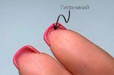
The question of why hyponychium appears is a strange one, to say the least, since the hyponychium of the nail (from Greek onychos - nail + hypo - below, below) is the area of epithelium located between the nails and the skin of the fingertips.
We can say that the hyponychium is located under the nail, more precisely, under its free (distal) edge, which is formed when the nail plate extends beyond the transition point between the skin of the finger and the nail, where the hyponychium fixes the nails at the ends of the fingers.
Also, the hyponychium - along with the onychodermal tract (the nail isthmus in the distal part of the nail bed at the transition to the hyponychium) - is a barrier that seals the subnail space and protects it from water, chemicals and microorganisms. [1]
What does hyponychium look like?
Delimiting the transition from the nail bed to the epidermis of the finger, hyponychium has the appearance of soft tissue thickening under the free edge of the nail plate. The epidermis of hyponychium is thickened, 90-95% consisting of keratinocytes; there is also a granular (granular) layer, in the cytoplasm of the cells of which there are granules of keratogialin - the initial protein for the formation of keratin. The outer, horny layer in the distal part (closer to the nail bed) is compact, and closer to the free edge of the nail plate - orthokeratotic (thicker) with keratinocytes, which are able to mature from mitotic to terminally differentiated state and replace dead cells. The underlying dermis (without subcutaneous tissue) is located directly on the last (distal) phalanx of the finger.
If you look at the top of a healthy nail, the hyponychium is not very visible, but if you look under the nail from the palm side, if you look closely, you can see a tiny strip of skin securing the nail to the end of the finger.
By the way, periungual skin structures (skin rolls) include paronychium, eponychium, and hyponychium. Paronychium is a skin roll that frames the edges of the nail plate. Eponychium is the proximal fold of skin that forms the cuticle (the thin horny layer on the nail plate). The cuticle and eponychium form another seal of the nail bed.
How does a hyponychium grow?
In the 11th week of gestation, a group of fetal cells migrate from the proximal nail furrow and spread proximally into the fingers, differentiating into nail matrix rudiments. And a ridge appears in the distal half of the nail field, which eventually differentiates into the hyponychium. The emergence of nail plates from under the proximal nail shaft is fixed at the 13th week of intrauterine development, and by the 32nd week the fetal nail unit already consists of the nail plate, nail matrix, nail bed, eponychium and hyponychium.
Normally, the hyponychium grows only to the transition point between the skin of the finger and the nail.
Causes of the hyponychia
For overgrowth of the epithelium between the skin of the fingertips and the part of the nail plates protruding above it, wording such as: overgrown hyponychium, large or protruding hyponychium, and enlarged or thickened hyponychium may be used.
When the epidermis at the transition between the skin of the finger and the nail protrudes above the pad of the finger up the back of the nail plate, the hyponychium is said to be longer than the nail.
The following causes can cause hyponychia overgrowth:
- Nail trauma;
- Growing long nails (with a much longer free edge of the nail plates), as well as frequent manicures with gel nail extensions or wearing acrylic nails for long periods of time;
- Dermatophyte-induced fungal nail disease - onychomycosis, especially distal and lateral subnail fungus (which first affects the hyponychium and then spreads to the nail plate and nail bed);
- Simple or allergic contact dermatitis;
- Acrodermatitis persistent pustularis allopo, which is often caused by localized trauma or infection of the last phalanges of the fingers;
- Nail psoriasis;
- Subnail hyperkeratosis leading to thickening of the nail plates - pachyonychia;
- Pitting or papular palm and plantar keratoderma;
- Syndrome or reiter's disease.
Risk factors
Among the risk factors for hyponychia overgrowth are noted leading to maceration of the skin prolonged contact with water, exposure to chemicals or nail polish and nail strengthening products, the presence of subnail and periungual formations (Warts, osteochondroma, exostosis, glomus tumor of the subnail space, fibrokeratoma, epidermal onycholemmal cysts, etc.); onychogryphosis (thickening of the nail and its deformity in the form of a bird's claw).); onychogryphosis (thickening of the nail and its deformity in the form of a bird's claw).
In addition, there are genetically determined features of the nails and periungual skin structures, in particular, such congenital (or acquired due to nail trauma, subnail exostosis or hyperkeratosis) pathology as pterygium inversum unguis - inverse or inverse nail pterygium. In this pathology, the hyponychium attaches to the underside of the nail as it grows, and the distal part of the nail bed fuses with the inner surface of the nail plate.
Pathogenesis
In cases where hyponychia overgrowth occurs due to frequent manicures with gel nail extensions or long-term wear of acrylic nails, the mechanism is explained by increased stress on the distal free edge of the nail plate, to which the hyponychia epithelium responds by activating cell division. And the longer the free edge of the nail, the more mechanical stress affects the subnail area.
In nail psoriasis, as in subnail hyperkeratosis, the proliferation and differentiation of cells of the stratum corneum is impaired; thickening and destruction of the nail is observed.
In the case of fungal nail infection, the pathogenesis of hyponychium damage is due to deformation of the nail plate and thickening of the nail bed skin, which causes the nail to lift and the hyponychium epithelium to peel away from the underlying tissues.
Symptoms of the hyponychia
Damaged hyponychium often leads to separation of the nail from the bed - onycholysis.
Patients with nail plate psoriasis or subnail hyperkeratosis often notice that the hyponychium has detached or the hyponychium has moved away from the nail.
The hyponychium, like the skin of the fingertips, has a lot of sensitive nerve endings, and the hyponychium (or rather, the entire fingertip) hurts, for example, when the fingers are burned, dermatitis or persistent acrodermatitis. If somehow the hyponychium is torn off - most often it happens with a traumatic nail tear, the acute pain can be unbearable.
With swelling, redness and pain, the inflammation of hyponychia becomes apparent, e.g. In the case of hyponychia panaricia, fungal nail infections or inflammatory onychopathy such as retronychia - with the voluminous formation of granulation tissue under the nail plates.
Diagnostics of the hyponychia
Hyponychia thickening can affect one, a few or all of the nails - depending on the cause.
To detect an overgrown hyponychium, a simple examination by a dermatologist or podologist is not enough: it is difficult to assess the condition of the hyponychium with the naked eye, so onychoscopy - dermatoscopy of the nails - is used. [2]
And to find out the cause of its overgrowth, a differential diagnosis is carried out.
Who to contact?
Treatment of the hyponychia
What is the treatment for hyponychia overgrowth? It is necessary to treat the disease that caused it. For example, in onychomycosis, antifungal drugs are used: nail fungus drops, as well as ointments and creams for nail fungus.
Dermatitis is treated with ointments and creams for dermatitis, nail psoriasis uses corticosteroids and non-hormonal ointments for psoriasis.
If hyponychia overgrowth is provoked by gel nail extensions or acrylic nails, the problem is eliminated by refusing these procedures.
How to remove hyponychium under the nails? It should not be removed: it is an integral anatomical part of the nail unit and a protective barrier of the subnail space.
How to grow hyponychium? It is not necessary to regrow it: as mentioned above, keratinocytes of its thickened stratum corneum are able to mature and replace dead cells.
What happens if the hyponychium is torn off? First of all, it will be very painful, and secondly, the natural protective barrier of the subnail space will be broken, with the threat of infection getting there.
What should I do if I damage the hyponychium? Leave it alone, as its epithelium has the potential to regenerate. [3]
Prevention
To avoid problems with hyponychium, you need to:
- Take good care of your nails;
- Avoid traumatizing your fingernails and fingertips;
- Do not grow long nails and do not abuse their extension, as well as a long time not to "weight" their nail plates with acrylic "prostheses". See - top 5 dangers of nail extensions;
- Protect your nails (and skin) from chemicals (there are gloves for this);
- Treat nail fungus and all dermatologic diseases and other pathologic conditions that can cause problems with the nails and peri-nail skin structures.
In conclusion, what hyponychia is. In terms of terminology, it is based on the Greek onychos - nail and the prefix hypo-, which also indicates the lowering of something below normal (for example, in the terms "hypovitaminosis", "hypotonia", etc.). And the question of why hyponychia occurs is more than appropriate, since this rare, usually congenital nail pathology or anomaly is defined as the absence of a part of the nail (half nail hypoplasia) or the presence of a rudimentary nail.
This pathology may be a genetic trait or the result of impaired nail formation during intrauterine development. Hyponychia - often in combination with skeletal anomalies - is mostly found in syndromes caused by mutations in genes encoding structural proteins of the skin and its appendages.