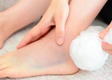
Dislocation of the foot: causes, symptoms, diagnosis, treatment
Last reviewed: 23.04.2024

All iLive content is medically reviewed or fact checked to ensure as much factual accuracy as possible.
We have strict sourcing guidelines and only link to reputable media sites, academic research institutions and, whenever possible, medically peer reviewed studies. Note that the numbers in parentheses ([1], [2], etc.) are clickable links to these studies.
If you feel that any of our content is inaccurate, out-of-date, or otherwise questionable, please select it and press Ctrl + Enter.

Dislocations in the ankle joint, as a rule, are combined with fractures of the ankles or anterior and posterior margins of the tibia. Isolated dislocations of the segments of the foot or its individual bones are relatively rare.
 [1]
[1]
Subtalar dislocation of foot
ICD-10 code
- S93.0. Dislocation of the ankle joint.
- S93.3. Dislocation of another and unspecified part of the foot.
Dislocation occurs at the level of the talus-calcaneus and talon-navicular joints from excessive indirect violence. Most often, as a result of excessive flexion and internal rotation of the foot, a dislocation of it occurs posteriorly with supination and internal rotation. However, when changing the direction of violence, dislocations of the foot are possible anteriorly, outwardly and internally.
Symptoms of subtalar dislocation of the foot
Pain is typical . Deformation of the foot depends on the type of displacement. With posterior-internal dislocations, the anterior part of the foot is shortened. The foot is biased towards the inside and back, supined and maximally bent. The talus bone will stand on the outer surface.
Diagnosis of subtalar dislocation of the foot
The final diagnosis is made after radiography.
Conservative treatment of subtalar dislocation of the foot
General anesthesia. To eliminate the dislocation proceed immediately upon diagnosis. Procrastination can lead to the formation of bedsores in places of pressure protruding bones and due to rapidly increasing edema.
The patient is placed on his back, the leg is bent to a 90 ° angle in the knee and hip joints. Fix the lower leg. The foot is further displaced towards the dislocation and tracts along the axis of the displaced segment. The second stage creates a counter-support in the standing bone, the foot is returned to the correct position. When the correction is heard click and appear movements in the ankle joint. Impose a posterior trough-like deep longe from the ends of the fingers to the middle third of the thigh for 3 weeks. With moderate edema, you can impose a circular bandage for the same period, but immediately cut it along the length and squeeze the edges. Flexion in the knee joint should be 30 °, in the ankle - 0 °. After 3 weeks, replace the gypsum bandage with a circular dressing, shortening it to the upper third of the shin. The period of immobilization is extended for another 8 weeks. Load on the limb in a plaster bandage is allowed no earlier than 2 months.
Estimated period of incapacity for work
The ability to work is restored in 3-3,5 months. During the year the patient should use the instep.
Dislocation of the ram bone
ICD-10 code
S93.3. Dislocation of another and unspecified part of the foot.
The mechanism of trauma indirect: excessive reduction, supination and plantar flexion of the foot.
Symptoms of a dislocation of the talus
Pain in the area of the injury, the ankle joint is deformed. The foot is tilted to the inside. A dense protrusion is felt along the anterior surface of the foot. The skin above it is whitish in color due to ischemia.
Diagnosis of talus dislocation
On the roentgenogram determine the dislocation of the talus bone.
Conservative treatment of talus dislocation
Elimination of dislocation is performed under anesthesia and immediately after diagnosis due to the danger of skin necrosis in the area of standing of the talus. The patient is laid in the same way as to eliminate the subtalar dislocation. They produce intensive traction for the foot, giving it even more plantar flexion, supination and reduction. Then the surgeon pushes the talus bone inside and back, trying to deploy it and move it to its own bed. The limb is fixed with a circular cast strip from the middle of the thigh to the ends of the fingers when bending at the knee joint at an angle of 30 °, in the ankle - 0 °. The bandage is cut lengthwise to prevent compression. After 3 weeks, the bandage is changed to a gypsum boot for a period of 6 weeks. After eliminating immobilization, rehabilitation treatment is carried out. To avoid aseptic necrosis of the talus, the load on the limb is allowed no earlier than 3 months after the injury.
Dislocation in the joint of a Sopar
ICD-10 code
S93.3. Dislocation of another and unspecified part of the foot.
Dislocation in the talon-navicular and calcaneocuboid joints occurs with a sharp deflection or leading (often abduction) rotation of the forefoot, which moves to the rear and to one side.
Symptoms of dislocation in the joint
Sharp pain, stop is deformed, edematous. The load on the limb is not possible. Circulatory circulation of the distal foot is disturbed.
Diagnosis of dislocation in the joint
On the roentgenogram, there is a violation of congruence in the joint of Chopar.
Conservative treatment of dislocation in the joint of the Chopar
Immediately and only under anesthesia eliminate the dislocation. Produce traction for the calcaneal region and forefoot. The surgeon eliminates the displacement of pressure on the rear of the distal foot and in the direction opposite to the displacement.
Apply a plaster boot with a well-modeled vault. The limbs are elevated for 2-4 days, after which they allow walking on crutches. The period of immobilization is 8 weeks, then impose a removable longure for 1-2 weeks, in which the patient walks on crutches with gradually increasing load. Further, rehabilitation treatment is carried out.
Estimated period of incapacity for work
Workability is restored after 12 weeks. The wearing of the instep for a year is shown.
Dislocation of foot in the joint of Lisfranca
ICD-10 code
S93.3. Dislocation of another and unspecified part of the foot.
Dislocations of metatarsal bones often arise from direct violence, often combined with fractures of the base of these bones. The displacement of the dislocated bones can occur outside, inside, in the rear or the plantar side.
Symptoms of dislocation of the foot in the joint of Lisfranca
Pain in the place of injury. The foot is deformed: shortened, thickened and widened in the anterior part, moderately supinated. The support function of the foot is broken.
Diagnosis of dislocation of the foot in the joint of Lisfranca
On the roentgenogram determine a dislocation in the joint of Lisfranc.
Conservative treatment of a dislocation of the foot in the joint of Lisfranca
The correction is carried out under general anesthesia. Assistants stretch the foot along the longitudinal axis, capturing the anterior and posterior parts along with the shin. The surgeon removes the existing displacement by the pressure of the fingers in the direction opposite to the dislocation.
The limb is immobilized with a plaster boot for 8 weeks. Give your leg an elevated position, assign a cold to the foot, control the state of the blood circulation. Circular gypsum bandage after the expiration of the period is removed and a removable gypsum longite is applied for 1-2 weeks. The load on the limb is allowed after 8-10 weeks.
Estimated period of incapacity for work
The ability to work is restored in 3-3,5 months. During the year, the wearing of the instep support is shown.
 [10], [11], [12], [13], [14], [15], [16]
Dislocation of toes
Of all the dislocations in the joints of the lower limb, only the dislocations of the toes are subject to outpatient treatment. The most frequent among them is a dislocation of the first finger in the metatarsophalangeal joint in the back.
ICD-10 code
S93.1. Dislocate the toe (s) of the foot.
Symptoms of dislocated toes
I finger is deformed. The main phalanx is located above the metatarsal at an angle that is open at the rear. Movement in the joint is absent. Mark a positive symptom of springing resistance.
Diagnosis of dislocation of toes
With the help of roentgenography, a dislocation of the first toe is detected.
Treatment of dislocation of toes
The method of correction is exactly the same as when removing the dislocation of the first finger of the hand. After manipulation, the limb is immobilized by a narrow back gypsum langetto from the lower third of the leg to the end of the finger for 10-14 days. Assign a subsequent recovery treatment.
Estimated period of incapacity for work
Workability is restored in 3-4 weeks.

