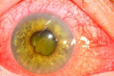
Iridocyclitis
Last reviewed: 23.04.2024

All iLive content is medically reviewed or fact checked to ensure as much factual accuracy as possible.
We have strict sourcing guidelines and only link to reputable media sites, academic research institutions and, whenever possible, medically peer reviewed studies. Note that the numbers in parentheses ([1], [2], etc.) are clickable links to these studies.
If you feel that any of our content is inaccurate, out-of-date, or otherwise questionable, please select it and press Ctrl + Enter.

Causes of the iridocyclitis
According to the etiopathogenetic trait, they are divided into infectious, infectious-allergic, allergic non-infectious, autoimmune and developing in other pathological conditions of the organism, including metabolic disorders.
Infectious-allergic iridocyclitis occurs against a background of chronic sensitization of the body to internal bacterial infection or bacterial toxins. More often infectious-allergic iridocyclitis develop in patients with metabolic disorders with obesity, diabetes, renal and hepatic insufficiency, vegetative-vascular dystonia.
Allergic non-infectious iridocyclitis can occur with drug and food allergies after blood transfusion, the introduction of sera and vaccines.
Autoimmune inflammation develops against the background of systemic diseases of the body: rheumatism, rheumatoid arthritis, children's chronic polyarthritis (Still's disease), etc.
Iridocyclitis can manifest itself as a symptom of a complex syndrome pathology: ophthalmostomatogenital - Behcet's disease, ophthalmourethrosinovial - Reiter's disease, neurodermatovouveitis - Vogt-Koyanagi-Harada disease, etc.
Pathogenesis
The inflammatory process in the anterior part of the vasculature can begin with the iris (iritis) or the ciliary body (cyclite). In connection with the generality of blood supply and innervation of these departments, the disease passes from the iris to the ciliary body and vice versa - iridocyclitis develops.
The above features of the structure of the iris and the ciliary body explain the high incidence of inflammatory diseases of the anterior segment of the eye. They can be of different nature: bacterial, viral, fungal, parasitic.
A dense network of wide vessels of the uveal tract with a slowed flow is practically a sedimentation tank for microorganisms, toxins and immune complexes. Any infection that develops in the body can cause iridocyclitis. The most severe course is inflammatory processes of the viral and fungal nature. Often the cause of inflammation is focal infection in the teeth, tonsils, paranasal sinuses, gall bladder, etc.
Symptoms of the iridocyclitis
Out of exogenous effects, the causes of development of iridocyclitis can be concussions, burns, injuries, which are often accompanied by the introduction of infection.
The clinical picture of inflammation distinguishes between serous, exudative, fibrinous, purulent and hemorrhagic iridocyclitis, acute and chronic in nature of the flow, focal (granulomatous) and diffuse (non-granulomatous) forms of inflammation in the morphological pattern. Focal pattern of inflammation is typical for hematogenous metastatic infection.
The morphological sub-extract of the main focus of inflammation in granulomatous iridocyclitis is represented by a large number of leukocytes, there are also mononuclear phagocytes, epithelioid, giant cells and necrosis zone. From such a focus can be identified pathogenic flora.
Infectious-allergic and toxic-allergic iridocyclitis occur in the form of diffuse inflammation. In this case, the primary damage to the eye can be outside the vascular tract and located in the retina or optic nerve, from where the process spreads to the anterior section of the vascular tract. In those cases when the toxic-allergic lesion of the vascular tract is primary, it never has the character of a real inflammatory granuloma, but arises suddenly, develops rapidly as a hyperergic inflammation.
The main manifestations are a violation of microcirculation with the formation of fibrinoid swelling of the vascular wall. In the focus of the hyperergic reaction there are edema, fibrinous exudation of the iris and ciliary body, plasma lymphoid or polynucleic infiltration.
Forms
- secondary uveitis;
- uveal keratitis - edema of the corneal stroma, folding of the decemet membrane, involvement of the sclera - keratosclerouveitis;
- Complicated (sequential) cataracts due to dystrophic processes in the lens, qualitative and quantitative changes in the intraocular fluid, as well as long-term administration of glucocorticoids;
- optic neuritis, which can lead to partial atrophy of the optic nerve;
- exudative and gracious retinal detachment;
- subatrophy and atrophy of the eyeball.
Complications and consequences
Outcome of iridocyclites:
- favorable with full recovery (normal corneal properties and visual functions are restored);
- slight bleaching of the cornea, pigment precipitates on the cornea and clouding of the lens, partial atrophy of the pupillary rim, pupillary deformity, destruction of the vitreous humor;
- complicated cataract; secondary uveitis
- atrophy of the eyeball;
- retinal disinsertion;
- cornea throat (if keratitis is attached).
The last three types of complications lead to a sharp decline in vision, up to blindness.
What do need to examine?
How to examine?


 [
[