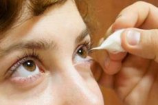
Assessment of the sensitivity of the cornea
Last reviewed: 23.04.2024

All iLive content is medically reviewed or fact checked to ensure as much factual accuracy as possible.
We have strict sourcing guidelines and only link to reputable media sites, academic research institutions and, whenever possible, medically peer reviewed studies. Note that the numbers in parentheses ([1], [2], etc.) are clickable links to these studies.
If you feel that any of our content is inaccurate, out-of-date, or otherwise questionable, please select it and press Ctrl + Enter.

Indications for assessing the sensitivity of the cornea
- Diseases of the cornea.
- Neuro-ophthalmologic pathology.
Method and interpretation of the corneal sensitivity assessment study
Fingers of the left hand dilute the patient's eyelids, gently touch the end of the cotton wick first center of the cornea, and then four points on its periphery. With normal sensitivity, the patient marks a touch or tries to close the eye. If this does not happen, then the thicker parts of the wick begin to be laid on the cornea. The appearance of the corneal reflex when laying a thicker part of the wick indicates a significant decrease in the sensitivity of the cornea. If this method fails to cause the corneal reflex, then there is no sensitivity.
Alternative methods for assessing the sensitivity of the cornea
A more accurate definition of the sensitivity of the cornea is carried out with the help of graduated hair by Frey-Samoilov. The sensitivity of the cornea is measured at 13 points of the cornea by three hairs (0.3: 1.0 and 10.0 g / mm squared). Algezimeters are also used, but the most advanced devices at the present time are optoelectronic esteziometers.


 [
[