Trichinella
Last reviewed: 23.04.2024

All iLive content is medically reviewed or fact checked to ensure as much factual accuracy as possible.
We have strict sourcing guidelines and only link to reputable media sites, academic research institutions and, whenever possible, medically peer reviewed studies. Note that the numbers in parentheses ([1], [2], etc.) are clickable links to these studies.
If you feel that any of our content is inaccurate, out-of-date, or otherwise questionable, please select it and press Ctrl + Enter.

Trichinella spiralis (Trichinella spiralis) - a worm of the class of nematodes (Enoplea), the family Trichinelloidea, living in the body of vertebrates carnivores - is pathogenic to humans. The disease caused by this helminth is called trichinosis.
According to infectious disease parasitologists, trichinella is found on all continents except Antarctica, and cases of systemic trichinosis have been documented in 55 countries. Trichinosis is considered one of the most serious and dangerous food zoonotic diseases caused by parasitic organisms. The death rate due to infection with Trichinella is 0.2-8%.
Structure of the trichinella
Trichinella is a relatively small round worm: the length of adult females ranges from 2.5 to 3.5 mm; male - from 1.2 to 1.8 mm; body diameter - 36 microns. The form of Trichinella Spiralis (as the name suggests) is spiral, and worms can twist and unwind, especially active in the anterior part of the body, which is conical and round.
The skin-muscular body of the worm is covered with a thin hypodermis, and on top is a strong cuticle consisting of the fibrillar collagen protein, which is a buffer against the host's immune response. In the head part of the adult nematode there is a mouth cavity with a protruding acute process (stiletto), which passes into the esophagus (and then into the three-stage intestine with digestive glands in the muscular walls).
Nematodes of Trichinella spiralis have sensory organs: motion detecting bristles (mechanoreceptors) and amphides of chemical detection (chemoreceptors).
Larvae of Trichinellae (0.08 mm long and up to 7 μm in diameter) are covered with a two-layered membrane, the inner layer has a large number of very thin fibrils located parallel to the larval circumference. Outside there is a pointed ledge.
The trichinella reproduces sexually in the small intestine, in the wall of which adult individuals live about 4-6 weeks. During this time, one female worm produces up to 1-1.5 thousand larvae. Then adult Trichinella perishes and is excreted from the body with feces.
The egg in the body of the female is fertilized by the sperm of the male. Each fertilized egg develops into a whole celluloid, which in the course of morphogenetic changes is transformed into a larval-fetus (trophocyte). Larvae of Trichinella fill the uterus of the female worm and after 5-6 days they leave it. Further they penetrate into the mucous membrane of the small intestine, and from it into the lymph and blood, spreading throughout the body. So the migratory phase of the larval invasion begins.
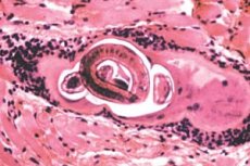
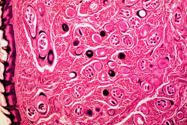
It should be noted that only larvae survive to the striated muscles, since only cells of skeletal muscle tissue can support the existence of the parasite. The larva not only hides in such cells from the host's immune system, forming a collagen capsule, but also stimulates the development of blood vessels around the affected cell in order to obtain the necessary nutrients.
In the protective cyst passes the first larval - infectious stage Trichinella; here the anaerobic larva can be from 15 days to several months or dozens of years, preserving viability in capsules that become calcified and acquire the appearance of intramuscular cysts.
Life cycle of the trichinella
The ways of infection with trichinella are only food, that is, the parasite enters the human body through the consumption of meat of animals infected with pathogenic larvae encased in capsules-cysts. In gastric juice, the capsules dissolve, and the larvae freely penetrate into the intestinal mucosa, where they - in the course of several lines - develop into adult worms.
The life cycle of a trichinella occurs in the body of one host (animal or human), and the worm does not need to go outside. The development and colonization of Trichinella spiralis occurs during four larval and one adult stages. The first stage of the larva passes in the striated musculature, and in the mucosa of the small intestine - three subsequent larval stages (which represent the molting process) and the stage of the adult worm. The immature small Trichinella feeds on the contents of the mucous cells, damaging them with styling, and in 3-4 days is ready for reproduction.
Thus, the life cycle of a trichinella begins with the enteral phase of infection, when a person or animal eats contaminated meat containing the first stage larvae - muscle.

The place of localization of trichinella is characteristic: chewing striated muscles of the head; oculomotor muscles of the orbit and orbit of the upper jaw; diaphragmatic muscles, skeletal muscles of the shoulder, neck and lumbar region. Perhaps this is due to a high level of vascularization of these muscle groups, as well as a significant myoglobulin content in the sarcoplasm of the membranes surrounding skeletal muscle cells.
Pathogenesis
Invasion of the larva through the intestine and its path to muscle tissue causes the pathogenic effect of trichinella.
First, the movement of the larva, "paving" its way to the right place, is accompanied by the inevitable destruction of cell membranes, loss of cytoplasm and damage to organelles, which causes cell death.
Secondly, the migration of newborn larvae with blood and lymph flow can enter them not only in the tissues of striated muscles, but also in cells of the liver, kidneys, lungs, myocardium and brain. And the more larvae "wander" through the human body in search of a suitable place in the muscles, the more severe the results of the invasion. This is expressed in general edema, increased excretion of protein with urine (proteinuria), a violation of calcium metabolism in the body, cardiomyopathy and anomalies of the central nervous system.
Thus, the pathogenic effect of trichinella can lead not only to parasitic myositis with persistent pains, but also to life-threatening diseases such as myocarditis, encephalitis, meningitis, nephritis. Trichinella in children can cause eosinophilic pneumonia or bronchopneumonia, myocarditis, meningoencephalitis. Read more - Trichinosis in children
Symptoms
Clinical symptoms of trichinosis are largely correlated with the number of larvae that enter the body, with the stage of infection (enteral or muscular), as well as with the state of the human immune system. So the infection can be subclinical.
The initial symptoms of the enteral phase, which can manifest 24-48 hours after eating contaminated meat, include general malaise and weakness, fever and chills, hyperhidrosis, diarrhea, nausea and vomiting, abdominal pain, which are caused by intestinal invasion of larvae and adults worms. These symptoms are nonspecific and inherent in many intestinal disorders, so in many cases this phase of infection (lasting from two weeks to a month) is diagnosed as food poisoning or intestinal flu.
Manifestations of trichonella invasion can slowly increase, as larvae migrate through the lymphatic system to the muscles. On the background of intestinal symptoms appear cough, headaches, swelling of the face and eye area, conjunctival hemorrhage or retina, petechia under the fingernails, muscle pains, cramps, itchy skin and papular rashes. These symptoms can persist for up to eight weeks.
A severe degree of infection with trichinella can lead to impaired coordination of hand movements; loss of motor functions (including walking); difficulty in swallowing and breathing; weakening the pulse and lowering blood pressure; kidney dysfunction; development of inflammatory foci in the lungs, heart, brain; nervous disorders.
Forms
Nematodes of the genus Trichinella infect a wide range of mammals, birds and reptiles. In addition to Trichinella spiral (parasitizing in the organism of definitive hosts - domestic pigs and wild boars, other synanthropic and wild carnivores), there are such species of this helminth as: Trichinella nativa, found in polar bears, seals and walruses of the Arctic; Trichinella nelsoni - in African predators and scavengers; Trichinella britovi - in carnivorous Europe, West Asia and North-West Africa; Trichinella murelli - in bears, moose and horses in North America.
These species of trichinella, invading the cells of the host's muscle tissue, form collagen capsules around cells with worm larvae that ensure their safe development.
But trichinella pseudospiralis (Trichinella pseudospiralis), parasitic in mammals in temperate zones, having a morphological similarity with Trichinella spiralis, refers to non-encapsulating varieties. More often than not, trichinella pseudospiralis, as the main hosts, has predatory birds, including migratory birds, which broadens the geographic range of the parasite.
In addition, non-encapsulating trichinella include Trichinella papuae, a parasite of wild and domestic pigs and combed crocodiles in Papua New Guinea and Thailand, as well as infecting African reptiles Trichinella zimbabwensis.
Diagnostics
Early clinical diagnosis of trichinella is quite difficult, because pathognomonic signs are absent. In addition, the diagnosis during the first week of infection is complicated by the fact that an increase in the synthesis of enzymes of creatine phosphokinase (CK) and lactate dehydrogenase (LDH), found in a blood test, is noted in other infections.
The levels of eosinophilic granulocytes in the blood serum also increase, but it is also non-specific for trichinosis and may indicate other parasitic infections, allergies or the presence of a malignant tumor in the patient.
The presence of Trichinella larvae in the body is indicated by antibodies to trichinella (IgG, IgM and IgE), which can be detected in the patient's blood as early as 12 days after infection - with serological examination of the blood sample using indirect immunofluorescence and latex agglutination. More information in the article - Trichinosis analysis: antibodies to Trichinella spiralis in the blood
There is an opportunity to identify DNA Trichinella by PCR, but the cost of such a study is too high for most hospital laboratories.
Diagnosis of infection with Trichinella also involves a muscle biopsy, for which a tissue sample is taken from the deltoid muscle. But with an insignificant number of larvae encapsulated in muscle tissue and a 17-24 day incubation period, the response of this study may be false-negative.
So indirect evidence of infection with this parasite can serve as bilateral periorbital edema, pinpoint hemorrhages under the nail plates, as well as high temperature combined with the history of eating undercooked meat.
Treatment
According to specialists, treatment of trichinella with anthelminthic drugs is possible only at an early stage of infection, while the parasite is in the small intestine. It is very difficult to drive out the larvae from muscle tissues by the current medicament means.
Nevertheless, an anthelminthic drug such as Albenzadol (other trade names Zentel, Gelmadol, Nemozol, Sanoxal) is prescribed - one tablet (400 mg) during meals, for 7-10 days. Also, the treatment of trichinella is carried out with Mebendazol (Vormin), which takes 2-4 tablets (0.2-0.4 g) three times a day for the first three days of treatment, and 0 for the next 7 days, 5 g (5 tablets each).
Systemic corticosteroids, in particular prednisolone, are also used simultaneously to prevent the aggravation of inflammatory reactions associated with the accelerated elimination of endotoxins (the so-called Yaris-Gerxheimer reaction). And muscle pain in trichinellosis is removed with the help of NSAIDs.
Alternative remedies for trichinella
Known helminthic alternative remedies for trichinella will not help if the parasite's larvae are already in muscle tissues. And on the enteric stage of trichinosis it is recommended to take decoctions of medicinal plants:
- a hundred-thousandth of an umbrella and an elephant (10 g of each herb per 200 ml of boiling water) - drink a few sips during the day;
- flowers chamomile pharmacy, tansy ordinary, grass cuffs sparkling and rhizome valerian - mix a tablespoon of each herb, a tablespoon of the resulting plant mixture, pour 250 ml of boiling water, boil for 10 minutes, insist under the lid for half an hour; Take 100 ml twice a day for 3-5 days.
And to relieve the inflammation of the intestines with diarrhea, you need to use the grass seedgrass, grass ivan-tea (kapreya izkolistnogo), sporisha (mountaineer bird) and veronica officinalis. A mixture of herbs and decoction of it are prepared, as in the previous recipe.
Prevention of the trichinella
The main prevention of infection with trichinella is to consume high-quality meat that has passed sanitary and veterinary control, use caution to consume game, and also subject meat to long-term heat treatment. It should be borne in mind that smoking, quick frying (steaks with blood), steaming or in the microwave oven do not kill Trichinella larvae: the meat should be cooked at a temperature of + 70-75 ° C, and it is safer to cook it longer.
Increased precautions require the use of pork. Parasitologists recommend freezing pork at -20 ° C for 7-10 days (or at -15 ° C for three weeks) to neutralize this parasite. In this case, the thickness of a piece of meat should not exceed 10 cm.
For the prevention of trichinella, an important veterinary control is very important when growing livestock for meat. In the EU countries, by the decision of the European Commission, since 2005, every batch of meat supplied by the producers is being tested for larvae of Trichinella spiral.


 [
[