Medical expert of the article

New publications
Zygomycetes are the causative agents of zygomycosis
Last reviewed: 06.07.2025

All iLive content is medically reviewed or fact checked to ensure as much factual accuracy as possible.
We have strict sourcing guidelines and only link to reputable media sites, academic research institutions and, whenever possible, medically peer reviewed studies. Note that the numbers in parentheses ([1], [2], etc.) are clickable links to these studies.
If you feel that any of our content is inaccurate, out-of-date, or otherwise questionable, please select it and press Ctrl + Enter.
Zygomycoses (phycomycoses) are caused by zygomycetes, which are lower fungi with non-septate hyphae (fungi of the genera Rhizopus, Absidia, Rhizoinucor, Basidiobolus, Conidioboius, Canninghameila, Saksenaea, etc.).

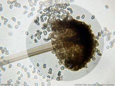
Morphology and physiology of zygomycetes
Zygomycetes consist of hyphae without partitions. Reproduction is asexual with the formation of sporangiospores and sexual with the formation of zygospores. From the spore-bearing hyphae - sporangiophores, rounded sporangia depart, containing spores and pus pores. Zygospores are formed during the sexual process and as a result of the fusion of two cells that have not differentiated into gametes. The aerial mycelium of some zygomycetes (Rhizopus) has arcuately curved hyphae - "whiskers" or stolons. Mycelium is attached to the substrate by special branches.
Zygomycetes antigens
Antigens of fungi are different: Mucor mucedu forms large (up to 200 µm) yellow-brown sporangia with oval spores; Rhizopus nigricans forms dark-brown mycelium with blackening sporangia (up to 150 µm in diameter) containing rough spores; Absidia cotymbifera forms sporangia 40-60 µm in diameter containing colorless ellipsoid, smooth, and less often rough spores. Zygomycetes are aerobes. They grow on simple nutrient media, Sabouraud medium; optimum growth is at 22-37 °C.
Pathogenesis and symptoms of zygomycosis
Fungi cause mycoses in people with weakened immunity. They produce lipases and proteases, which facilitate the spread of fungi in tissues. In immunodeficient people, fungi penetrate the blood vessels, causing thrombosis. A fulminant form of infection is known - rhinocerebral zygomycosis. Ischemic tissue necrosis and the formation of a polymorphonuclear infiltrate occur. A distinction is made between invasive pulmonary zygomycosis, as well as gastrointestinal and skin forms of the disease. The brain, eyes and other organs and tissues are also affected. Patients develop cellular immunity, accompanied by DTH.
 [
[