New publications
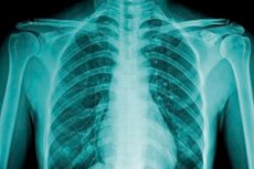
X-ray of the thoracic spine in two positions
Last reviewed: 29.06.2025

All iLive content is medically reviewed or fact checked to ensure as much factual accuracy as possible.
We have strict sourcing guidelines and only link to reputable media sites, academic research institutions and, whenever possible, medically peer reviewed studies. Note that the numbers in parentheses ([1], [2], etc.) are clickable links to these studies.
If you feel that any of our content is inaccurate, out-of-date, or otherwise questionable, please select it and press Ctrl + Enter.
The spinal column is the most important part of the musculoskeletal system. Its condition determines the smooth functioning of almost all organs and systems. There are many methods of diagnosing the spine, but most often the choice of doctors stops at radiography. In our article, we will consider one of the most common types of research - X-ray of the thoracic spine, which allows us to assess the condition of the vertebrae, characterize them, and determine biomechanical features.
Indications for the procedure
The doctor may recommend an X-ray of the thoracic spine if it is necessary to diagnose or follow the dynamics of such pathologic conditions:
- Diseases affecting bone tissue and cartilage (osteochondrosis, intervertebral hernias, spondylosis, spondyloarthritis);
- Neurological symptoms;
- Traumatic back injuries (if vertebral injuries are suspected);
- Congenital or acquired deformities of the spinal column (kyphosis, scoliotic curvature, pathologic lordosis).
In addition, the doctor may resort to X-rays if the patient complains of discomfort in the back - especially related to physical activity (bending, turning, etc.).
X-rays of the thoracic spine are often recommended when these symptoms are present:
- Unpleasant sensations (pain, crunching, numbness, tingling, etc.) in the upper back or upper extremities;
- Mechanical damage to the spinal column, intervertebral hernias, suspected tumor processes;
- Curvature of the thoracic spine;
- Chest pain, difficulty breathing, heart pain.
The doctor may require an x-ray of the thoracic spine if necessary:
- Evaluate the vertebrae for misalignment, damage, shape changes, and vertebral spacing;
- Exclude inflammatory, degenerative and other processes in the spinal column;
- Consider the correct shape of the spine.
Preparation
The preparatory stage for X-ray of the thoracic spine is simple and involves following the rules listed below.
During 2-3 days before the diagnostic procedure it is desirable to follow some changes in diet, which is necessary to reduce gas formation in the intestine and correct display of the result on the image. You should exclude the use of whole milk (fresh fermented milk products are allowed), black bread, raw white cabbage, peas, dried fruits. In addition, alcohol and carbonated drinks should not be consumed.
If the diet was not observed, or the patient suffers from diseases of the digestive system, then he can follow the following recommendations: if flatulence a day before the X-ray of the thoracic spine should take a few tablets of any sorbent (even activated charcoal will do) or a preparation based on simethicone.
If the patient has excessive nervous excitability, it is recommended to start taking valerian or motherwort 2-3 days before the procedure.
X-ray of the thoracic spine should preferably be performed on an empty stomach. A light snack is allowed, without overeating (including on the eve of the study).
Technique of the x-rays of the thoracic spine.
X-rays of the thoracic spine can be performed in multiple projections:
- In front;
- The back one;
- Sideways.
The diagnostic process itself is not very difficult. The patient frees the upper part of the body from clothing, removes all metal accessories (jewelry, chains, watches, etc.). Then takes a position depending on the doctor's recommendations (standing, sitting, lying on the side or back). In some cases, it may be necessary to perform an image of the thoracic spine with the torso tilted forward.
The number of images and projection is agreed upon in advance with the doctor. Usually no more than 3-5 images are taken. The total time of the thoracic X-ray procedure is up to fifteen minutes.
At the time of imaging, the patient must remain still, because the quality of the images depends on it. During movements, the picture is "blurred", which significantly complicates diagnosis and may require repeated X-ray of the thoracic spine.
The results of the examination can be ready within an hour after the procedure. The X-ray image is first evaluated by a radiologist and then by the attending physician or a specialized specialist (vertebrologist, neurologist, surgeon, etc.).
X-ray of the thoracic spine with functional tests
Radiography of a particular section of the spinal column with functional tests - that is, with the performance of special exercises and shifting the center of gravity - is performed to more thoroughly determine the condition of the spine and its functional capacity. This helps to clarify the diagnosis and assess the severity of painful disorders.
Functional tests are additional conditions in which the condition and structural features of the spinal column can be examined in detail. Most often, the doctor asks the patient to assume a certain posture in which the spine is bent or extended at a certain angle. In this way, for example, it is possible to consider the displacement of the vertebrae in relation to each other, as well as the degree of their curvature. The procedure, like X-rays in general, is painless and lasts up to 15 minutes.
In addition to mechanical tests, it is possible to use X-ray contrast and drug tests. The first option involves examination of the blood network. Drug tests help to determine the structure and functionality of certain organs, such as the intestines, bronchi, esophagus, and so on.
Currently, functional tests are an effective way to obtain the most accurate information about the state of the body. However, such tests are not performed in relation to the thoracic spine due to inexpediency, examining only functional disorders of the cervical and lumbar spine, as well as extremities.
Contraindications to the procedure
When can an X-ray of the thoracic spine be contraindicated? Modern X-ray equipment makes this type of diagnosis extremely safe. The amount of radiation exposure is as low as possible and is almost comparable to the natural background. Specialists believe that there are no absolute contraindications to the X-ray of the thoracic spine. Nevertheless, this procedure can be carried out exclusively on the prescription of the doctor.
Temporary contraindications may include pregnancy (especially the first trimester) and early infancy (if the child cannot sit still for a few minutes).
Contrast radiography is not recommended in hypersensitivity to iodine preparations, thyroid pathologies, decompensation of diabetes mellitus, active tuberculosis.
Sometimes a thoracic X-ray can be difficult to perform - for example, if the patient is obese or mentally ill, or has metal fixed implants.
In general, the age or gender of the patient does not play a major role in performing an X-ray of the thoracic spine. Of course, the procedure should not be performed if there are no indications for it.
Normal performance
Today, radiography is used in a wide variety of medical fields - primarily because of the availability and informative nature of this method. X-ray of the thoracic spine allows to diagnose:
- Bone integrity disorders, tumor processes, arthritis, arthrosis, scoliosis;
- Tuberculosis, inflammatory processes;
- Cystic, polyposis and other benign masses;
- Anomalies and malformations;
- Salt deposits (calcinosis, etc.).
X-ray of the thoracic region is considered to be one of the most informative diagnostic procedures, allowing to clearly determine the zonality of the affected area. It is possible to assess the condition of the thoracic segment as a whole, or each vertebra individually.
X-rays help to determine the shape and structure of the thoracic vertebrae and intervertebral discs. It can detect deformities and other abnormalities of the spinal column.
In addition, with the help of X-ray of the thoracic region it is possible to detect such pathologies:
- Altered bone configuration and structure;
- Disrupted positioning of individual vertebrae;
- Deformity and lack of mobility of the spine;
- Bone disease;
- Disruption of the discs between the vertebrae;
- The presence of metastases;
- Osteoporosis.
X-ray of the thoracic region is considered mandatory after surgical interventions performed to restore normal spinal function.
Thoracic osteochondrosis on x-ray
If osteochondrosis of the thoracic is suspected, an X-ray of the spine is performed to check for changes in the height of the intervertebral discs, the presence of marginal overgrowths, deformity of the vertebral bodies, and indirect signs of herniation. These signs can be found in the vast majority of people over 50 years of age.
X-rays, as they pass through tissues and organs, are trapped in denser tissues. It is this property that makes it possible to thoroughly assess the condition of the skeletal bones and, in particular, the thoracic spine. Osteochondrosis is a painful process that at the very beginning of development affects the intervertebral discs. Then there are violations in the vertebrae themselves - bodies and ramifications, which is perfectly visualized in the course of radiography. The most characteristic radiological signs of osteochondrosis are bone overgrowth and narrowing of the intervertebral disc.
Thoracic osteochondrosis is accompanied by the separation of intervertebral discs, which in the course of reducing their own height begin to impinge on the intercostal nerves. Nevertheless, the symptomatology of the defeat of the thoracic department is noted quite rarely, which is due to the physiology of rib connections. With osteochondrosis, the upper thoracic vertebrae are more often affected, which are deformed. The clinical picture manifests itself only with age, against the background of dehydration of intervertebral tissues. Frequent trauma, physical overload, chronic intoxication aggravate the picture.
Due to the vagueness and late onset of symptoms, osteochondrosis is often detected accidentally, for example, when an MRI or X-ray of the thoracic spine is performed. Most often, reduced stability between the vertebrae - so-called segmental instability - is noted. To clarify the diagnosis, the doctor may recommend contrast radiography.