Medical expert of the article

New publications
Venous pulse and venous pressure.
Last reviewed: 04.07.2025

All iLive content is medically reviewed or fact checked to ensure as much factual accuracy as possible.
We have strict sourcing guidelines and only link to reputable media sites, academic research institutions and, whenever possible, medically peer reviewed studies. Note that the numbers in parentheses ([1], [2], etc.) are clickable links to these studies.
If you feel that any of our content is inaccurate, out-of-date, or otherwise questionable, please select it and press Ctrl + Enter.

The venous system supplies blood to the right heart. Therefore, when the pressure in the right atrium increases, corresponding to the increase in central venous pressure, due to heart failure, the peripheral veins dilate (swell), primarily the visible veins in the neck.
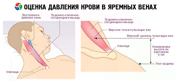
Normally, this pressure does not exceed 10 cm H2O and increases with right ventricular failure of any nature (especially with tricuspid valve defects, constrictive pericarditis and cardiac tamponade ). By swelling of peripheral veins, for example, the hand, one can roughly estimate the central venous pressure. Distinct swelling of the veins of the hand occurs when it is positioned at the level of or below the left atrium. If the hand is raised to a horizontal level above the left atrium, especially higher than 10 cm, a decrease in the blood filling of its veins is clearly noticeable. The vertical distance between the angle of Louis and the left atrium is on average 5 cm. By carefully moving the hand and observing the state of its veins, one can thus roughly estimate the central venous pressure.
Measuring venous pulse
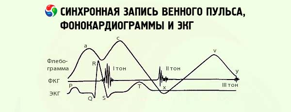
When recording the pulsation of the jugular vein, a curve is obtained that largely reflects the contractile function of the right chambers of the heart. The curve of the venous pulse consists of three positive waves. The highest of them, wave "a", precedes the main wave of the arterial pulse and is caused by the systole of the right atrium. The second wave c corresponds to the systole of the ventricles and is the result of the transmission of pulsation from the carotid artery. The third positive wave "v" is caused by the filling of the right atrium and, accordingly, the jugular vein during the closure of the tricuspid valve. When the tricuspid valve opens, a diastolic descent is noted on the curve of the venous pulse, since at this time the blood rushes from the atria into the right ventricle. This descent continues until the next wave.
The normal venous pulse is called atrial (or negative) because during the period when the arterial pulse curve descends (the lowest segment), the venous pulse curve has the greatest rise. During atrial fibrillation, the "a" wave disappears. The venous pulse can begin with a high "v" wave and turns into the so-called ventricular (or positive) venous pulse. It is called positive because the rise of the venous pulse curve is noted almost simultaneously with the main wave on the sphygmogram. A positive venous pulse is noted in case of tricuspid valve insufficiency (with an intense blood flow from the right ventricle to the atrium and veins).
Measuring venous pressure
Measuring venous pressure can also provide additional information about the condition of the peripheral veins of the neck and blood circulation in the systemic circulation. It is performed using a phlebotonometer, which is a glass tube with a lumen diameter of 1.5 mm with millimeter divisions from 0 to 350. The lower end of the rubber tube system is connected to a needle. The system of glass and rubber tubes is filled with a sterile isotonic sodium chloride solution. The fluid level in the sterile tube is set at the zero division of the scale. The subject is in a lying position. The device is positioned so that the zero division of the scale is located at the level of the right atrium, approximately at the lower edge of the pectoral muscle. The pressure is measured in the ulnar vein, into which a needle connected to the rubber tube of the device is inserted. In this case, the pressure in the vein and in the tube system is equalized. In healthy people, it fluctuates within 60-100 mm H2O. Its increase is noted in heart failure with blood stagnation in the systemic circulation.
The study of peripheral circulation, primarily arterial pulse, arterial pressure, and the condition of the veins of the neck, is important primarily for assessing the function of the heart. Along with this, local circulatory disorders associated with vascular diseases (both arteries and veins) and detected by conventional physical examination methods are possible.