Medical expert of the article

New publications
Uncomplicated fractures of thoracic and lumbar vertebrae
Last reviewed: 05.07.2025

All iLive content is medically reviewed or fact checked to ensure as much factual accuracy as possible.
We have strict sourcing guidelines and only link to reputable media sites, academic research institutions and, whenever possible, medically peer reviewed studies. Note that the numbers in parentheses ([1], [2], etc.) are clickable links to these studies.
If you feel that any of our content is inaccurate, out-of-date, or otherwise questionable, please select it and press Ctrl + Enter.
Uncomplicated compression wedge fractures of the lumbar and thoracic vertebrae are perhaps the most common type of spinal injury and are localized in the upper lumbar and lower thoracic spine.
What causes uncomplicated wedge compression fractures of the thoracic and lumbar vertebrae?
These vertebral body injuries occur as a result of the flexion mechanism of violence. By their nature, they are considered stable injuries.
The opinion of some authors that minor wedge-shaped compression of the vertebral bodies is completely harmless and is easily compensated by changing the position of the above and below parts of the spine is incorrect.
Often, even very minor compression of the vertebral bodies in the transitional lumbar-thoracic region, where these injuries most often occur, in the long term leads to severe complications in the form of pain syndrome and compression of the anterolateral sections of the spinal cord. The cause of these complications are progressive degenerative changes in adjacent intervertebral discs, aggravated by a previous injury and the seemingly minor deformation of the vertebral body that has arisen.
These seemingly harmless “minor” fractures of the vertebral bodies require the most serious attention.
Symptoms of compression fractures of the vertebral bodies
The most frequent and typical complaint is the presence of pain. Usually the pain is strictly localized at the level of injury, and increases with movement. Sometimes the pain is diffuse and spreads to the lumbar and thoracic regions. The pain syndrome is most pronounced in the first hours and days after injury, and at a later stage it significantly smooths out and even disappears.
The pain is most distinct and pronounced when the victim is in a vertical position while walking. Its intensity increases when walking on uneven ground, when driving in a car, etc. Often these pains are accompanied by a feeling of uncertainty in the "strength of the spine", and discomfort.
Diagnosis of compression fractures of the vertebral bodies
A detailed examination of the anamnestic data, the circumstances of the injury and the place of application of violence allows us to suspect the presence of a wedge-shaped compression fracture of the vertebral bodies and its probable localization.
 [
[ Inspection
Often the victims are quite active. The degree of the existing deformation of the spine is sometimes so little expressed that it is detected only by an experienced eye. In the lumbar region, this deformation may manifest itself only by smoothing out the physiological lordosis, against which a button-shaped spinous process is visible in thin subjects. Often, this protrusion of the spinous process is determined only by palpation. In the thoracic region of the spine, some increase in physiological kyphosis is determined, against which a button-shaped protrusion of the spinous process is more clearly visible. In addition to the deformation of the spine in the sagittal plane, there may also be a lateral curvature of the line of the spinous processes, indicating the presence of lateral compression of the vertebral body.
A slight spinal deformity may be masked by the existing swelling of soft tissues at the fracture level. This swelling is absent in the first hours after the injury and appears later.
When examining the victim, it is almost always possible to detect tension in the long muscles of the back, determined by eye, limited to the area of injury, or spreading to the entire lumbar and thoracic spine. Sometimes, topical muscle tension is determined only by palpation, especially in subjects with pronounced subcutaneous tissue.
Palpation reveals local pain at the level of the spinous process of the fractured vertebra. In the later post-traumatic period, in the presence of kyphotic deformation, local pain is determined at the level of the spinous process of the vertebra located above the fractured vertebra. Palpation reveals an increase in the interspinous space, which is more pronounced the greater the compression of the body of the fractured vertebra. Palpation can also reveal a spinal deformity that was not detected during examination.
The pain symptom with axial load on the spine is usually not detected in the lying position. It is not so valuable that it is necessary to give the victim a vertical position to detect it, since this position is not always safe for the victim.
Spinal mobility
Many authors note the limitation of the volume of active movements in case of spinal injuries. There is no doubt that, as with any injury to the musculoskeletal system, there is a limitation of the mobility of the spine in case of its injury. However, this method of examining the victim in the presence of acute spinal injury should be excluded from clinical practice as unjustified and risky for the victim.
Of particular interest is the examination of active movements in the legs. As is known, with uncomplicated spinal injuries, active movements in the legs are preserved. If you ask a victim with a compression wedge fracture of the vertebral body in a supine position to bend at the hip joints and slightly spread the legs straightened at the knee joints, then pain always occurs in the area of the fracture. This pain symptom persists much longer than others.
Thompson's symptom can help in diagnosing an uncomplicated compression wedge fracture, which consists in the fact that pain in the spine at the level of injury in a sitting position disappears when the spine is unloaded by the victim's hands resting on the seat of a chair.
Other clinical symptoms observed in uncomplicated compression wedge fractures of the bodies may include reflex urinary retention, pain in the posterior abdominal wall upon deep palpation, arising due to the presence of a retroperitoneal hematoma.
Sometimes, for the same reason, there is tension in the anterior abdominal wall, sometimes so pronounced that it simulates the picture of an “acute abdomen”, but for which a laparotomy is performed.
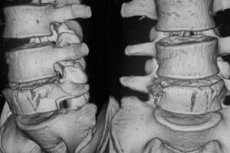
Spondylography
The X-ray examination method is one of the most important and in many cases decisive addition to the clinical examination in case of compression wedge fractures of the vertebral bodies. Spondylography is performed in two typical projections - posterior and lateral. The lateral spondylogram is decisive in making a diagnosis.
Compression wedge fractures of the vertebral bodies are characterized by typical radiological symptoms that allow not only to confirm or reject the suspected clinical diagnosis, but also to clarify and detail the existing damage.
The most typical radiographic symptom is the wedge-shaped form of the vertebra with the apex of the wedge facing neutrally. The degree of this wedge-shapedness is highly variable - from controversial, barely perceptible, to absolutely indisputable, well-defined and striking. Collapse, some thickening and especially rupture of the ventral endplate make the diagnosis of a fracture indisputable. These data are determined on a profile spondylogram: change and unevenness of the bone structure of the vertebral body, displayed on spondylograms (direct and lateral) by thickening of the bone beams of the vertebral bodies along the compression line; rupture of the endplate, more often the cranial one, of the vertebral body. In the thoracic region, damage to the cranial endplate is often stepwise; when the endplate, more often the cranial one, is ruptured, a lateral spondylogram shows its indentation and disruption of continuity (acute Schmorl's node). rupture of the cranioventral angle of the vertebral body, revealed on the profile spondylogram; narrowing of the intervertebral space and the area of adjacent intervertebral discs, more often in the ventral sections; an increase in the interspinous space, determined on the anterior and lateral spondylograms; axial deformation of the spine, more often in the sagittal, less often in the frontal plane. In case of lateral compression of the vertebral body, a wedge-shaped deformation of the body cannot be detected on a profile spondylogram, but it is possible to detect compaction of the bone structure of the body at the cranial endplate. In these cases, an anterior spondylogram allows us to determine lateral compression of the body. In case of compression fractures of the thoracic vertebrae, a paravertebral hematoma is formed due to significant bleeding, which on the anterior spondylogram forms a fusiform paravertebral shadow resembling an abscess.
In some cases, spondylography in oblique projections may be useful. With a minor degree of compression and the absence of distinct radiographic symptoms of a vertebral body fracture, it is not always possible to radiologically confirm the clinical diagnosis of the existing injury. In these cases, it is recommended to repeat the radiographic examination after 6-10 days. By this time, due to bone resorption along the fracture line, its display on the X-ray film becomes more distinct.
Based on clinical and radiological data, in typical cases it is not difficult to recognize and diagnose a compression wedge fracture of the lumbar and thoracic vertebral body. Spondylography allows to clarify and detail the nature of the injury, its features and shades. Serious difficulties may be encountered in recognizing mild, insignificant degrees of compression of the vertebral bodies, especially in the thoracic region. Additional spondylograms, including targeted ones, and sometimes tomographic examination, analysis of clinical and radiological data in dynamics in the absolute majority of cases allow to get closer to the truth.
In the presence of relevant clinical and anamnestic data indicating a vertebral body fracture, in the absence of convincing radiographic symptoms, one should lean towards the diagnosis of a fracture and treat the victim as having a vertebral body fracture. Only when convincing and indisputable evidence of the absence of damage subsequently appears can one abandon the presumptive diagnosis. Such tactics will protect the victim from unwanted and sometimes severe late complications that arise in the case of undiagnosed damage.
Treatment of uncomplicated compression wedge fractures of the bodies of the thoracic and lumbar vertebrae
In the treatment of uncomplicated compression wedge fractures of the thoracic and lumbar vertebral bodies, as in the treatment of fractures in general, the ultimate goal is to restore the anatomical shape of the damaged segment and restore its function. There is no doubt that, more often than not, restoration of the anatomical shape of the damaged bone segment, with proper treatment, contributes to a more complete restoration of function. Unfortunately, this seemingly obvious position is most often violated in the treatment of uncomplicated compression wedge fractures of the vertebral bodies. Many traumatologists have a firmly rooted idea that the loss of the correct anatomical shape of the body of one vertebra does not pose any problems for the victim and is easily compensated for by changing the position of other segments of the spinal column. It is this concept that is one of the main reasons for unsatisfactory movements, which are not so rarely observed in the treatment of these injuries.
The ideal method of treating uncomplicated compression wedge fractures of the lumbar and thoracic vertebral bodies is one that would restore the anatomical shape of the damaged vertebral body, eliminate vertical load on it, reliably maintain the position of the achieved reclination and create long-term immobilization of the damaged segment of the vertebra for the period necessary for healing of the fracture, without limiting the function of the above and below parts of the spine. The generally accepted existing methods of treating compression wedge fractures of the vertebral bodies do not meet all these requirements. The method we propose using a "tie" fixator is not ideal in the full sense of the word.
Among the existing methods of treating uncomplicated compression wedge fractures of the lumbar and thoracic vertebrae, the main ones are:
- method of one-stage repositioning followed by immobilization with a plaster corset;
- gradual stage reposition method;
- functional method;
- surgical treatment methods;
- complex functional method using a clamp-type device.
Method of one-stage reposition with subsequent immobilization with a plaster corset. The expediency and possibility of restoring the anatomical shape of the body of a broken vertebra by extension and hyperextension of the spine was expressed by Henle at the end of the 19th century. The implementation of this idea in practice was restrained by the fear of possible damage to the spinal cord during repositioning. In 1927, Dunlop and Parker demonstrated in practice the possibility of restoring the anatomical shape of a broken vertebra by stretching and extending the spine. Wagner and Stopler (1928) succeeded in achieving straightening of the body of a broken vertebra in a number of victims, but failed to maintain it in the position of the achieved correction. Only after 1929, when the works of Davis were published, and subsequently Boliler, Watson Jones, B. A. Petrov, I. E. Kazakevich, A. P. Velikoretsky and others, a detailed developed and substantiated method of one-stage reposition entered everyday practice. In our country this method has not become widespread.
One-stage reduction is performed under local anesthesia using the Shneck method. The victim is placed on his side. By palpation, focusing on local pain, in comparison with the spondylography data, the spinous process of the damaged vertebra is determined. In case of damage to the lumbar vertebra, stepping back 6 cm from the line of the spinous processes to the side on which the victim is lying, mark the point of needle insertion. A 16 cm long injection needle is inserted through the wetted point from the bottom up at an angle of 35°. As the needle advances, the tissue is anesthetized with a 0.25% solution of novocaine. Depending on the severity of subcutaneous fat and muscles, at a depth of approximately 6-8 cm, the tip of the needle rests against the posterior surface of the transverse process. The injection needle is pulled back slightly, its angle of inclination is changed in no way so that when moving in depth it slides along the upper edge of the transverse process. At a depth of 8-10-12 cm, the tip of the needle rests against the posterolateral surface of the body of the broken vertebra. 5 ml of 1% novocaine solution is injected with a syringe. The syringe is removed from the needle pavilion. If blood-stained liquid is released from the needle pavilion, this means that the needle has been inserted into a hematoma in the area of damage. Otherwise, the needle is removed and reinserted according to the method described above one vertebra higher or lower. No more than 10 ml of 1% novocaine solution should be injected into the area of the broken vertebra to avoid complications in the event of a puncture of the dura mater or penetration of novocaine through a possible rupture into the subarachnoid space.
When anesthetizing the body of a thoracic vertebra, the injection needle is inserted at the level of the spinous process of the overlying vertebra, since the spinous processes of the thoracic vertebrae are located more vertically and their apices are below the corresponding body.
Anesthesia of the fractured vertebral body can also be achieved by injecting 40 ml of 0.25% novocaine solution into the interspinous space between the damaged and adjacent vertebra. Once in the hematoma, the anesthetic solution reaches the fracture area. Anesthesia of the fractured vertebra can also be achieved by intraosseous anesthesia - by injecting 10-50 ml of 0.25% novocaine solution into the spinous process of the damaged vertebra. In this latter case, anesthesia is achieved for a very short time, since the novocaine solution is quickly carried away by the venous blood flow.
If the anesthesia is performed technically correctly, the pain in the area of the broken vertebra will disappear or decrease significantly rather quickly.
Simultaneous reduction technique
One-stage reduction can be achieved in various ways. Bohler performs one-stage forced reduction using two tables of different heights; they are placed in a line so that there is a gap between them that allows free access to the victim's torso along the lumbar and most of the thoracic spine. The victim is placed in a prone position so that his legs and lower torso are placed on the lower table, approximately up to the level of the anterior superior iliac spines. He rests on the higher table with his axillary areas and arms bent forward at the elbows. In this position, the victim's spine seems to sag between the tables and is "hyperextended."
The victim remains in this position for 15-20 minutes, after which a plaster corset is applied, which maintains the position of the spine achieved during the reclination process.
Watson Jones performs a one-stage forced reduction using traction through a block fixed to the ceiling. For this, the victim is placed on the table in a prone position. In case of damage to the lumbar vertebrae, traction is carried out with special straps for the lower parts of the shins of straightened legs, in case of damage to the upper lumbar vertebrae or lower thoracic vertebrae - with special straps for the rib cage. In the position of the achieved "hyperextension" a plaster corset is also applied.
The degree of achieved straightening of the body of the fractured vertebra during the forced one-stage reduction is monitored using profile spondylograms.
The question of the duration of wearing a corset after a single-stage forced reposition is very important. B. A. Petrov, Bohler consider a period of immobilization with a plaster corset of 2-3 months to be sufficient, I. E. Kazakevich, Watson Jones - 4-6 months, and Kazmirowicz (1959) - 8-9 months. It is well known that the healing process of the body of a broken vertebra is quite long and lasts 10-12 months. For this reason, external immobilization with a plaster and then a removable corset should be long-term - at least 1 year, otherwise secondary compression of the broken vertebra may occur. Wearing a plaster and removable orthopedic corset should be accompanied by therapeutic massage and gymnastics aimed at preventing the development of atrophy and weakness of the muscles.
The method does not pose any danger if it is used according to the correct indications only for uncomplicated wedge-shaped compression fractures of the bodies of the thoracic and lumbar vertebrae.
The main disadvantage of this method of treating compression wedge fractures of the vertebral bodies is the need for long-term wearing of a plaster cast, and then a removable orthopedic corset. The negative aspects of immobilization with a corset are well known. These include unhygienic conditions, the need to immobilize undamaged parts of the spine, which puts the spine in conditions of passive relaxation, limitation of the function of the chest and its organs, atrophy and weakness of the muscles. The most significant disadvantage of this method of treatment is the inability to quite often prevent secondary deformation of the body of the broken vertebra.
The method of staged repositioning of the broken vertebral body consists not in one-time, but in gradual, staged straightening. Various authors have proposed various devices in the form of pads, special frames, supports, etc.
The simplest and most effective method is the staged repositioning by A. V. Kaplan. It is as follows. Immediately upon admission to the hospital, the victim is placed on a hard bed in a supine position. A small, dense bolster is placed under the lower back. A day later, this bolster is replaced with a higher one, and after another 1-2 days, a large bolster 15-20 cm wide and 7-10 cm high is placed under the lower back. As a result of "hyperextension" on the bolster, the broken vertebra gradually straightens out and its anatomical integrity is restored. According to the author of the method, this method is easier for victims to tolerate - they gradually get used to the dosed "hyperextension", while intestinal paresis, urinary retention and other possible complications do not occur, or rather, occur less frequently. In some cases, the author advises combining staged straightening with one-time traction along an inclined plane. During the staged straightening of the broken vertebra, spondylography is used to control the body.
On the 8th-15th day, a plaster corset is applied for "small displacements" for a period of 2-3 months, and for "large" ones - for 4 months. Working capacity is restored in 4-6 months. Patients engaged in heavy physical labor are transferred to light work within a year from the end of treatment.
A. V. Kaplan (1967) notes that in recent years, after staged repositioning, he has been fixing broken vertebrae by the spinous processes with metal plates. This suggests that staged repositioning followed by long-term wearing of a corset does not always lead to favorable outcomes.
The functional method of treating uncomplicated wedge fractures of the lumbar and thoracic vertebrae has become especially widespread in our country. To this day, it is the method of choice for treating compression fractures of the vertebrae in many trauma hospitals.
The functional method is based on the concept of Magnus (1929, 1931) and Haumann (1930) that a compression wedge fracture of the body of a lumbar or thoracic vertebra is impacted, and this in itself promotes faster healing of the fracture and eliminates the possibility of secondary displacement, so straightening this vertebra is inappropriate and unlikely (Klapp). According to V. V. Gornnevskaya and E. F. Dreving, a plaster corset, delaying the regeneration of a broken vertebra and causing muscle atrophy, causes more harm than good.
Based on the above, the authors of the method believe that straightening the body of the broken vertebra is harmful and that there is no need to seek restoration of the anatomical shape of the broken vertebra during treatment. The main thing in treating this type of injury, in their opinion, is the creation of a good "muscle corset", which is achieved by therapeutic exercises; The authors believe that therapeutic exercises accelerate the regeneration processes in the broken vertebra, that under the influence of systematic "traction and dosed load" there is an appropriate restructuring of the spongy substance of the body of the broken vertebra and the bone trabeculae are located in statically advantageous directions during the restructuring process.
To create a “muscle corset”, E. F. Dreving developed a well-structured system of therapeutic gymnastics, including four periods.
The essence of the method is that the victim is placed on a hard bed with an inclined plane for traction using the Glisson loop and rings for the axillary areas. From the first hours and days, therapeutic exercises are started, aimed at strengthening and developing the muscles of the spine, back and abdomen. After 2 months, by the time the victim gets to his feet, a well-defined "muscle corset" is formed, holding the spine in a state of some hyperextension.
The functional focus of the method, its simplicity and accessibility, the lack of need for active manipulation and wearing a corset led to the fact that this method quickly gained significant distribution. The experience of its practical application for 35 years has revealed a number of significant shortcomings. These include the impossibility of maintaining the correct regimen during treatment. Thus, according to A. V. Timofeevich (1954), 50% of victims treated with the functional method did not maintain the required regimen and were discharged from the hospital early. Only 10% of victims performed the recommended outpatient treatment. This is explained by the fact that after the acute effects of the injury have passed, the victims feel healthy, forget about the spinal fracture and do not want to burden themselves with treatment. It is not always possible to form a "muscle corset" (especially in elderly and obese people, in weakened patients with concomitant diseases) . The disadvantage of the method is the need for a long stay in bed, etc. However, the most serious disadvantage of this method is the refusal to restore the anatomical shape of the broken vertebra, which, in our deep conviction, is the main cause of subsequent complications.
Surgical treatment methods
The surgical methods of treating spinal injuries described in the literature relate to the treatment of various other clinical forms of its injuries and are not directly related to the treatment of uncomplicated compression wedge-shaped fractures of the bodies of the lumbar and thoracic vertebrae. Only in recent years have some authors proposed methods of surgical treatment of uncomplicated compression wedge-shaped fractures of the bodies of the lumbar and thoracic vertebrae.
Complex functional method using a clamp-tie
A method that is close to the ideal for treating uncomplicated compression wedge fractures of the lumbar and lower thoracic vertebrae is one that would allow for reliable immobilization of the damaged segment of the spine after restoration of the anatomical shape of the broken vertebra for the period necessary for healing of the fracture, and at the same time would not interfere with the creation of a “muscle corset”, freeing the victim from the need to stay in bed and wear a corset.
The complex functional method of treatment with temporary internal fixation of the damaged section of the spine with a "tie" fixator, proposed by us and developed with the participation of E. A. Ramikh and A. I. Koroleva, meets some of the above-mentioned tasks. The basis of this method is temporary internal fixation of the damaged section of the spine with a special metal "tie" fixator.
The use of metal to fix broken vertebrae is not new. Wilkins (1886) was the first to tie broken vertebral arches with wire. Novak (1952) was the first to use wire sutures in the treatment of uncomplicated compression wedge fractures of the vertebral bodies in a group of victims. Havlin (1961) modified the technique of applying wire sutures. Ladio (1959) uses a fenestrated screw metal fixator to stabilize fracture-dislocations of the thoracic and lumbar localization.
Indications: closed uncomplicated compression wedge fractures of the bodies of the lower thoracic and lumbar vertebrae.
In the process of treatment, three periods are conventionally distinguished. The first period covers the period of time from the moment the victim is admitted to the hospital until the implementation of surgical internal fixation.
The goal of the first period is to eliminate the acute effects of the previous injury, improve the general condition of the victim, correct the axial deformation of the spine, and restore the anatomical shape of the broken vertebra.
This same period is preparatory for the subsequent internal fixation. Its average duration is 7-10 days.
Immediately upon admission of the victim to the hospital, establishment of the diagnosis and clarification of the location of the injury, anesthesia is administered to the site of injury.
Anesthesia of the fractured vertebral body is performed according to Shneck. The anesthesia technique is described above. The victim is placed on a hard bed. A fabric hammock is placed under the damaged section of the spine, with metal cables attached to the ends and thrown over blocks secured to two Balkan frames on the bed. A 3-5 kg load is suspended from the cables. During the first 3-5 days, the loads are increased to 12-18 kg, depending on the victim's weight. With the help of such gradual reclination, it is possible not only to correct the axial deformation of the spine, but also to restore the anatomical shape of the fractured vertebral body. Using a hammock for reclination is more convenient for both the patient and the staff than using sand bags or other hard reclinators.
From the second day, the victim begins to do therapeutic exercises according to the complexes developed by A. I. Koroleva and E. A. Ramikh. These gymnastic complexes are based on the method of E. F. Dreving, which is modified taking into account the short period of the patient's stay in bed and subsequent early gymnastics in a standing position. The first complex, designed for the first 2-3 days, mainly provides for general hygienic exercises. Much attention is paid to breathing exercises. At the same time, exercises designed to strengthen the back extensors are gradually included. At the end of the first period, exercises are introduced for more active training of the back and abdominal muscles, some strength exercises for the upper limbs, "half-scissors", and walking in place, etc.
The second period of complex functional treatment covers a short period of time necessary to perform internal fixation of the damaged section of the spine by surgery with a metal clamp.
The clamp-"tie" consists of a coupling sleeve and two hooks. The coupling sleeve is a cylindrical tube 50 mm long. Its internal diameter is 4.5 mm, external - 6 mm.
Anesthesia is usually performed by local layered infiltration with 0.25% novocaine solution and supplemented by the introduction of 1% novocaine solution to the body of the broken vertebra. It is quite acceptable, and in particularly reactive patients, endotracheal anesthesia is preferable. In these cases, muscle relaxation is performed at certain moments of the intervention. During this period, the patient is transferred to controlled breathing.
A universal surgical operating table is used, on which the victim is placed in a prone position.
Guided by anatomical landmarks, compared with the available anteroposterior spondylogram, the spinous process of the broken vertebra is localized, which is marked with a metal injection needle inserted into its apex. It should be borne in mind that it is not always easy and simple to determine the spinous process of the broken vertebra, since usually by the time of surgery the axial deformation of the spine is eliminated and the pain reaction to pressure disappears.
The technique of the internal fixation operation of the damaged section of the spine is as follows. The skin, subcutaneous tissue, and superficial fascia are dissected layer by layer by a median linear incision along the line connecting the tops of the spinous processes. The tops of the spinous processes covered by the supraspinous ligament are exposed. On the right or left, depending on the nature of the spinal deformation in the bridge of injury, the lumbosacral fascia is dissected at the lateral surface of the spinous processes 0.5 cm from the midline. The choice of the side of the fascia dissection, and ultimately the side of the installation of the "tie" fixator depends on whether there is an angular deformation of the spine to the side. If there is one, it is more advantageous to install the fixator on the convex side of the deformation; if there is no angular deformation, then it makes no difference on which side the fixator is installed.
The size of the skin incision is approximately equal to the length of 4-5 vertebrae. Using a scalpel, scissors and a spinal raspatory, the long muscles of the back are separated from the lateral surface of the spinous processes and arches partly sharply, partly bluntly, along the length of the fractured overlying and underlying vertebrae. The inevitable bleeding is stopped fairly quickly by tamponade with gauze napkins soaked in hot saline. The bases of three spinous processes and interspinous spaces filled with interspinous ligaments become visible in the wound.
One of the hooks of the clamp-coupler is unscrewed from the coupling. The hooks of the clamp-coupler, one of which is left in connection with the coupling, are inserted into the interspinous space with their sharp curved end, they cover the upper surface of the spinous process of the vertebra located above the broken vertebra. The coupling is placed at the base of the spinous processes along their lateral surface. The second hook, previously unscrewed, is inserted into the interspinous space with its tip, it covers the lower surface of the spinous process of the vertebra located under the broken vertebra, its end, bearing the thread, contacts the coupling. Usually three vertebrae are subject to fixation: the damaged, the upper and the lower one. Accordingly, the hooks of the clamp-coupler are installed. A control radiography is performed in the anteroposterior projection, with the help of which the surgeon is convinced that the clamp is inserted correctly.
Having made sure of the exact position of the clamp, the surgeon anesthetizes the area of the broken body by injecting 10 ml of 1% novocaine solution. Naturally, this manipulation is performed only if the intervention is performed under local anesthesia!
The patient is given an extension position. If a lumbar vertebra is broken, then a greater hyperextension is given to the foot end of the body; if the lower thoracic vertebra is damaged, then hyperextension is given to the head end of the body. This position is given to the patient with the help of a cable, secured with a leather cuff either on the shins of the victim or on the chest and the position of the operating table.
In the hyperextension position, the "tie" fixator twists and stabilizes the damaged section of the spine in the position of the achieved correction. If the compressed vertebra is not fully straightened, its body is further straightened by tensioning the fixator. In the hyperextension position, the main load of the overlying section of the spine falls on the posterior, undamaged section of the spine, which promotes faster healing of the fracture.
It should be known that when performing surgical intervention under local anesthesia, the hyperextension position given to the victim is quite unpleasant for him. Therefore, he should remain in this position for a minimum amount of time.
During the operation, careful hemostasis is performed. The surgical wound is sutured layer by layer. A rubber strip is inserted into the subcutaneous tissue for 24 hours. An aseptic bandage is applied.
After acquiring some skill with careful, consistent and meticulous execution of the operation, its implementation is not difficult and takes a minimum of time.
The third period of complex functional treatment is the longest. It begins actually from the moment of completion of the surgical intervention and ends upon the patient's recovery.
The goal of the third period is the earliest possible rehabilitation of the victim and his return to useful work.
The presence of a strong and reliable fixation of the damaged section of the spine, achieved with the help of a “tie” fixator, creates optimal conditions for active functional therapy, which promotes the fastest healing of the fracture and the creation of a “muscle corset”.
Due to reliable internal fixation of the damaged section of the spine, 14-16 days after surgery, the victim can be put on his feet and active therapeutic exercises can be performed in a standing position. The effectiveness of early therapeutic exercises in a standing position in the absence of functional limitations in the undamaged sections of the spine is quite obvious.
The patient is placed on a bed with a shield in a supine position. A hammock with weights at its ends of 3-5 kg on each side is placed under the back at the level of the damaged section of the spine. During the first postoperative days, the victim usually receives painkillers and antibiotics. If necessary, appropriate symptomatic treatment is administered.
From the first day after the operation, the victim begins to do therapeutic exercises. The set of gymnastic exercises on the 1st - 3rd day is designed for 10-15 minutes and consists of general hygienic and general strengthening exercises. These are mainly static and dynamic breathing exercises (full breathing, abdominal breathing according to I. M. Sarkizov-Sirazini). Exercises are selected strictly individually, taking into account the patient's condition.
On the 2nd day after the surgery, the victim is allowed to carefully turn on his side. The bandage is changed, the rubber drain is removed, the wound is revised. An aseptic bandage is applied.
On the 4th day after the operation, a set of exercises is introduced, designed to strengthen the muscles of the lower extremities and back extensors. Breathing exercises are continued. With these gymnastic exercises, the victim is gradually prepared for the transition from a horizontal to a vertical position. The set of exercises is designed for 15-20 minutes and is repeated 5-6 times during the day.
Starting from the 7th day, the third set of gymnastic exercises is introduced. This set provides for even more intensive training of the back and lower limb muscles. Additionally, exercises are included in the prone position. On the 8th-9th day, the stitches are removed. On the 4th-16th day, the victim is allowed to stand up. The gymnastic exercises of this period are combined into the fourth set. It usually begins with a series of exercises from the previous sets, after which the victim is transferred to a vertical position. On the first day, the victim usually gets used to the vertical position, stands by the bed, and tries to walk around the ward. Gymnastics ends with a series of dynamic breathing exercises in the lying position.
Three to four days after the victim has moved to a vertical position, gymnastic exercises are performed mainly from a standing position. In addition to the strength exercises of the previous complexes, exercises for the lower extremities and pelvis, and for the back extensors are included. Free walking and breathing exercises serve as rest between exercises. This fifth complex is designed for 35-40 minutes.
Usually by the end of the 3rd - beginning of the 4th week after the internal fixation surgery, the victim is discharged in good condition for outpatient treatment. At home, he continues to do therapeutic exercises, mainly from the fifth complex. The duration of the exercises is 30-40 minutes 3-4 times a day.
Approximately by the end of the 2nd month after the operation, work not associated with significant physical exertion is permitted. Subsequently, systematic, constant exercise therapy is highly desirable.
This is the general scheme of complex functional treatment of uncomplicated wedge-shaped compression fractures of the lumbar and lower thoracic vertebral bodies. Naturally, depending on the individual characteristics of the victim, the nature and location of the injury, age, etc., this scheme may vary.
The described complex functional method of treatment using a "tie" fixator is the method of choice in the treatment of various types of uncomplicated compression wedge-shaped fractures of the bodies of the lumbar and thoracic spine, in particular uncomplicated compression wedge-shaped fractures of the bodies of the lumbar and thoracic vertebrae with varying degrees of reduction in their height, uncomplicated compression wedge-shaped fractures of the bodies of the lumbar and thoracic vertebrae with a rupture of the cranioventral angle, compression fractures of the lumbar vertebrae with a rupture of the lamina interdigitata - the so-called penetrating fractures.
S. S. Tkachenko (1970) modified the clamp-"tie", called it "special", and changed the technique of its application. The modification of the "tie" consists in a slight change in the angle of inclination of the hooks. In our opinion, this somewhat reduces the possibility of its "work" on twisting. More serious objections are available regarding the technique of intervention recommended by S. S. Tkachenko. The hooks-"ties" are attached to the spinous processes, and to the semi-arch, for which the yellow ligaments are first peeled off, a "partial resection of a part of the arch" is performed near its root. The hooks-"ties" are inserted into the defects formed during the partial resection of the arches. Thus, foreign metal bodies are introduced into the lumen of the spinal canal, to the interruption of which the epidural tissue will certainly react. It is difficult to say what effect all these factors will subsequently have on the relationship of the spinal cord with the walls of the spinal canal.
The author's recommendations to fix not 3 but 4 vertebrae in the case of a fracture of one vertebral body are hardly justified.
Anterior spondylodesis in the treatment of closed uncomplicated, "penetrating" fractures of the thoracic vertebral bodies
Closed compression wedge fractures of the thoracic vertebral bodies occur with a flexion mechanism of violence. In cases of damage to the cranial or, less commonly, caudal endplate, the intervertebral disc is also damaged - such a fracture should be classified as a more severe "penetrating" fracture.
Compression fractures of the lumbar vertebrae with a rupture of the cranioventral angle are also essentially "penetrating". However, with these injuries, the powerful lumbar intervertebral disc either does not suffer, or its damage is subsequently compensated to a certain extent by cicatricial healing of the disc. In the thoracic region, the intervertebral discs are weak and, as a rule, their damage entails the subsequent occurrence of intervertebral osteochondrosis.
It is known that any pathological process in the anterior spine entails the development of kyphotic deformation. This is especially characteristic of the thoracic spine, the anatomical norm of which is moderate physiological kyphosis. As a rule, this kyphosis increases and takes on a pathological character after compression fractures of the bodies of the thoracic vertebrae. This occurs due to the almost inevitable secondary decrease in the height of the body of the broken vertebra. Some surgeons believe that wedge-shaped compression of one vertebra and even axial deformation of the spine do not affect its function and do not cause pathological phenomena. Our numerous observations do not confirm this. A relatively small wedge-shaped deformation of the body of only one vertebra, without gross axial deformation of the spine, can lead to pain, functional failure of the spine, and in some cases, disability.
Existing methods of treating these spinal injuries are not always able to prevent the occurrence of these pathological phenomena. Experience shows that even early posterior spondylodesis in these cases may be ineffective,
The indication for anterior thoracic fusion is “penetrating” compression fractures of the thoracic vertebral bodies in young patients.
The main objective of anterior spondylodesis is to maintain the normal height of the anterior section of the damaged segment of the spine, prevent secondary compression of the bodies of the damaged vertebrae and axial deformation of the spine, and prevent the development of intervertebral osteochondrosis in the damaged discs. The most favorable time for intervention in the absence of contraindications is 5-7 days after injury. Pain relief is endotracheal anesthesia with controlled breathing.
The victim is placed on the operating table on his left side and slightly turned onto his back. The right arm is extended upward. The left leg is bent at the knee and hip joints.
Surgical access. Preference should be given to right-sided transpleural access, but left-sided access can also be used if necessary. Depending on the level of damage, the access level is selected: for lower thoracic - the level of the IX rib, for middle thoracic - the level of the VI rib.
The skin incision is made along the corresponding rib from the paravertebral to the anterior axillary line. The skin, subcutaneous tissue, and superficial fascia are dissected layer by layer. The superficial leaflet of the periosteum is dissected along the rib to be resected. The rib is isolated subperiosteally and resected along the length from the neck to the anterior axillary line. The deep leaflet of the periosteum and the parietal pleura are dissected. The pleural cavity is opened and examined.
If intrapleural adhesions are present, they are separated by blunt or sharp dissection depending on their nature. The edges of the chest wound are spread apart using a screw retractor. The lung is displaced toward the root - the anterolateral surface of the thoracic vertebrae becomes visible and accessible for manipulation. The intercostal vessels passing along the anterior surface of the bodies of the thoracic vertebrae, branches of the large splanchnic nerve and intervertebral discs protruding in the form of ridges are visible through the translucent mediastinal pleura. The pulsating thoracic aorta is clearly visible along the left axial surface of the spine. On the right, closer to the posterior lateral surface of the bodies of the thoracic vertebrae, the azygos vein is visible. The damaged vertebra is easily identified by the decrease in the height of its ventral wall, by the narrowed discs or discs that have lost their characteristic ridge shape. Subpleural hemorrhage often helps with orientation.
If there is the slightest difficulty in localizing the area of damage, one should resort to control radiography with preliminary marking of the suspected site of damage with injection needles.
The mediastinal pleura is dissected by linear incisions along the long axis of the spine, slightly to the right of the splanchnic line.
The incision of the mediastinal pleura should be made to the right of the midline in order to avoid conflict with the thoracic duct. The mediastinal pleura is peeled apart to the sides. If necessary, the aorta, left lateral surface of the vertebral bodies and left paravertebral region can be approached from the right-sided approach. After the incision of the mediastinal pleura, the anterior longitudinal ligament and the structures lying on it are exposed. The intercostal arteries and veins passing along the anterior surface of the vertebral bodies are isolated, ligated and dissected. The branches of the great splanchnic nerve are isolated and retracted to the lateral surfaces. The anterolateral surface of the vertebral bodies, the anterior longitudinal ligament and the intervertebral discs are exposed. The length of the exposure of the anterior surface of the spine depends on the number of damaged vertebrae.