Medical expert of the article

New publications
Types of pleuropneumonias
Last reviewed: 04.07.2025

All iLive content is medically reviewed or fact checked to ensure as much factual accuracy as possible.
We have strict sourcing guidelines and only link to reputable media sites, academic research institutions and, whenever possible, medically peer reviewed studies. Note that the numbers in parentheses ([1], [2], etc.) are clickable links to these studies.
If you feel that any of our content is inaccurate, out-of-date, or otherwise questionable, please select it and press Ctrl + Enter.
Pneumonia is classified according to the degree of lung damage. If the inflammatory process affects only the lobes, without spreading to the vessels and alveoli, then we speak of pleuropneumonia, or lobar pneumonia - an infectious disease that can be caused by viruses, microbes or fungi. In turn, there are different types of pleuropneumonia, which can only be identified by a medical specialist.
Today, there are a number of pleuropneumonias that differ in certain features. Such classification is necessary, first of all, for the optimal choice of treatment for the disease.
Forms
The division of various types of pleuropneumonia is based on clinical, etiological and other signs. For example, aspiration, post-traumatic, postoperative pleuropneumonia, as well as viral, bacterial, fungal, etc. are distinguished. Let's consider the basic types of pleuropneumonia, their features and main characteristics.
Infectious pleuropneumonia
Many types of pleuropneumonia differ depending on the infectious agent. Identification of the infection is mandatory, since the treatment regimen and the methods and drugs used depend on it. Infectious pleuropneumonia is classified as follows:
- Viral pleuropneumonia is caused by viruses and can be a complication of improper treatment or non-treatment of influenza or acute respiratory viral infections. Less often, it is a primary infection. It is diagnostically difficult to identify the virus in pleuropneumonia, so antiviral agents with a broad spectrum of activity are most often prescribed for treatment, as well as various symptomatic drugs.
- Mycoplasma pneumonia occurs after a special type of microorganism called mycoplasma penetrates the lung tissue. This disease is most often recorded in childhood and adolescence. It can proceed latent, without specific symptoms, but responds well to treatment with antibacterial drugs.
- Fungal pneumonia and pleuropneumonia can be caused by various types of infection, including fungal pathogens. Fungal pleuropneumonia is diagnosed only after a full diagnosis, since the clinical symptoms of this type of disease are usually scanty, the signs are blurred and unclear, and often do not correspond to the classic manifestations of microbial damage. The disease can be caused by mold fungi, Candida, endemic dimorphic fungi, pneumocysts. Most often, the "culprit" is Candida albicans, as well as aspergilli or pneumocysts - that is, an infection focused on the lung tissue. Pathogens can enter the respiratory system from both external foci and other mycotic foci present in the human body. For example, Candida is a permanent component of the skin and mucous microbiocenosis, but under certain circumstances it can be activated and become pathogenic: as a result, pneumomycosis develops. Treatment for fungal infection in the lungs is long-term, using a powerful antifungal course.
- Actinobacillosis pleuropneumonia is caused by Actinobacillus, a gram-negative capsule-forming pleomorphic rod. This disease affects only ruminants: cattle, pigs, and, less often, sheep. Other animals and humans are immune to the infection and do not get sick. Previously, before 1983, the disease was called "hemophilus pleuropneumonia": at the moment, this term is considered obsolete, since the pathogen, which was previously classified as belonging to the genus Haemophilus, has now been moved to the genus Actinobacillus.
Another predominantly veterinary term is "contagious pleuropneumonia." This refers to a particularly contagious form of pneumonia that is easily transmitted from one animal to another, causing widespread disease. The causative agent is usually Mycoplasma mucoides. Animals that have recovered from contagious pleuropneumonia become immune to the infection.
Abscessing pleuropneumonia
When speaking about abscessing pleuropneumonia, we mean the presence of foci of infectious purulent-necrotic destruction of the lung. These are multiple purulent-necrotic areas of tissue decay, and there is no clear boundary with healthy lung tissue. Due to the presence of characteristic destructive processes, many specialists call this disease the term "destructive pleuropneumonia".
In the lungs, zones of tissue melting of the confluent type are formed. The main causative agent of the pathology is considered to be Staphylococcus aureus, but there is also damage by Klebsiella and other enterobacteria, as well as hemolytic streptococcus, pneumococcus and anaerobic microbes.
The most common cause of the development of abscessing pleuropneumonia is considered to be aspiration of oropharyngeal secretions and the presence of foci of purulent infection inside the body adjacent to the lymphatic and blood vessels.
The symptoms of the disease are similar to those of total pneumonia.
Community-acquired pleuropneumonia
Community-acquired pleuropneumonia is one of the types of inflammatory pulmonary processes in which the infectious agent enters the respiratory system outside of a hospital or other medical and preventive institution. This form of pleuropneumonia can be bacterial or viral, and the transmission route is airborne.
In most patients, the inflammatory reaction is triggered after an untreated acute respiratory viral infection or influenza infection, tracheitis or bronchitis.
The pathogen enters the lungs via a descending route – from the upper respiratory tract. If the immune defense is weakened, it becomes difficult for the body to overcome new inflammatory foci. As a result, the infection settles in the lung tissue, and acute pleuropneumonia develops.
Often, patients with community-acquired pleuropneumonia already have various chronic respiratory processes, such as chronic bronchitis. The disease becomes active when certain conditions are created, when the immune system is weakened. If treatment is delayed or ignored, pleuropneumonia may develop.
Hypostatic pneumonia
A special form of the disease is hypostatic pleuropneumonia, which is predominantly secondary. Most often, the disease develops as a result of prolonged stagnation of blood circulation in the small circulatory system, which should provide trophism of the lung tissue. Impaired blood flow leads to the accumulation of intoxication products in the lungs. Viscous sputum is formed, to which microorganisms actively multiply - usually streptococci and staphylococci, which causes a new inflammatory process.
Hypostatic or congestive pleuropneumonia usually appears in patients who have been lying down for a long time and are unable to move and lead a normal life due to injuries or somatic pathologies. Thus, primary diseases can be heart attacks, strokes, diabetes, oncopathologies, etc. A prolonged horizontal position worsens blood flow and causes tissue congestion.
Types of pleuropneumonia depending on the extent of the lesion
The right lung is divided into three lobes, the left – into two lobes. In turn, each lobe is divided into segments – parenchymatous zones ventilated by a segmental bronchus and a certain branch of the pulmonary artery.
When the inflammatory reaction is located in one pulmonary lobe, it is called lobar pleuropneumonia, and in both lobes, it is called bilobar pleuropneumonia. Unilateral and bilateral lobar pleuropneumonia are also distinguished. The clinical picture and treatment measures are similar to other types of the disease.
In addition, specialists have identified the following types of lobar pathology:
- segmental pleuropneumonia – characterized by damage to one segment of the pulmonary lobe;
- polysegmental pleuropneumonia - indicates damage to several lobar segments at once;
- Upper lobe pleuropneumonia can be either right- or left-sided and indicates damage to the upper lobe of the lung;
- lower lobe pleuropneumonia can also be right or left-sided, depending on the localization of the pathological process;
- middle lobe pleuropneumonia is an inflammatory process in the middle lobe of the right lung (in the left lung the middle lobe is absent);
- total – occurs with damage to the entire lung field (all lobes of both the right and left lung);
- subtotal pleuropneumonia - for this form, damage to both lobes of one lung is typical;
- focal pleuropneumonia indicates a clear localization of the inflammatory focus, without spreading to nearby tissues;
- subpleural pleuropneumonia is an inflammatory process localized in the subpleural region of the lung;
- basal pleuropneumonia - characterized by an inflammatory reaction in the lower part of the lung.
This classification is based on the extent of the inflammatory reaction. The severity of symptoms depends on the extent of the lesion: the more extensive the inflammation, the deeper and more vivid the clinical picture. [ 1 ]
Confluent pleuropneumonia
In the confluent form of pleuropneumonia, painful disorders affect several areas of the lung at once, or even a pulmonary lobe. There is a pronounced lag in the breathing process on the affected side, and the symptoms of respiratory failure (shortness of breath, cyanosis) increase.
Confluent pleuropneumonia is characterized by infiltrative changes, against the background of which there are compacted infiltration zones and (or) destructive cavities. The term "confluent" means the fusion of multiple or single small pathological foci into larger formations. Given this feature of the development of pleuropneumonia, it is considered by specialists as a relatively unique form of the pulmonary inflammatory process.
Complications and consequences
If treatment measures were prescribed in a timely manner, and the treatment itself was competent, then the course of pleuropneumonia usually loses its typical cyclicity and is interrupted at the initial stage of development.
If the process of exudate resorption is disrupted, complications of pleuropneumonia develop. In some cases, connective tissue grows in the pathological focus: carnification occurs with subsequent pulmonary cirrhosis. In some patients, purulent processes with destruction (melting) of tissues are observed, and pleuropneumonia develops into an abscess or gangrene of the lung.
In pleuropneumonia, there are manifestations of the dry form of pleurisy with fibrinous layering and formation of adhesions. Lymphogenic spread of infection leads to the development of purulent mediastinitis and pericarditis. If the spread of microbes occurs through the circulatory system, then
Metastatic purulent foci in the brain and other organs and tissues: the development of purulent meningitis, peritonitis, acute polypous-ulcerative or ulcerative endocarditis, purulent arthritis begins.
Patients are often concerned about why the temperature does not drop while taking antibiotics for pleuropneumonia: could this indicate the development of complications? With pleuropneumonia, temperature readings usually fluctuate within 37-38°C. With antibiotic therapy, high temperature can persist for 2-3 days, and with a bilateral pathological process - up to 10-14 days (while not exceeding 38°C). If the readings exceed the limit of 39-40°C, this indicates an increase in the inflammatory reaction and the loss of the body's ability to fight the pathogen. In such a situation, the doctor should immediately review the treatment and, possibly, change the antibiotic. [ 2 ]
Diagnostics pleuropneumonias
Examination of a patient with suspected pleuropneumonia is carried out according to an individual plan drawn up by a doctor. Typically, this plan includes:
General blood, urine, sputum tests, blood biochemistry (determination of total protein, protein electrophoresis, determination of bilirubin and fibrinogen content);
Sputum culture with determination of bacterial flora sensitivity to antibiotic therapy;
ECG.
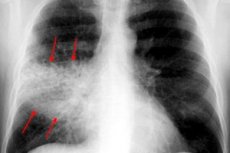
Chest X-ray is almost always the basic diagnostic method for all types of pleuropneumonia. The examination is performed in two projections:
- during the high-tide stage, an intensification and enrichment of the pulmonary pattern is observed, which is explained by tissue hyperemia;
- the degree of transparency is normal or slightly reduced;
- there is a uniform shadow, and the pulmonary root is slightly expanded on the affected side;
- if the pathological reaction is localized in the lower lobe sector, then a reduced excursion of the corresponding diaphragmatic dome is observed;
- During the hepatization stage, a marked decrease in the transparency of the lung tissue is detected (in accordance with the affected area);
- the affected area of the lung is of normal or slightly enlarged size;
- the intensity of the shadow increases slightly towards the periphery;
- in the middle zones of darkening, areas of lightening are found;
- the pulmonary root on the affected side is widened and has a uniform shadow;
- compaction of the adjacent pleura is observed;
- during the resolution stage there is a decrease in the intensity of the shadow of the pathologically altered zone;
- the fragmented shadow is reduced, the pulmonary root is expanded.
If there is a suspicion of pleuropneumonia, it is preferable to conduct a full X-ray examination, rather than standard fluorography, which is considered a preventive rather than a therapeutic and diagnostic method. Pneumonia is not always properly traced on fluorography, since this depends on both the severity of the pathological process and the condition and density of the tissues through which the X-rays penetrate. With the help of fluorography, it is possible to prevent the development of chronic pneumonia in advance, protect against an atypical course of the inflammatory process, however, this procedure does not allow you to note the localization of inflammation and assess the degree of complexity of the process.
Patients with any type of pleuropneumonia are recommended to have their external respiratory function examined, and if indicated, a pleural puncture is performed.
Multispiral CT is indicated in the following cases:
- if there are obvious clinical symptoms of pleuropneumonia, but there are no typical abnormalities on the X-ray image;
- if during the diagnosis of pleuropneumonia atypical disorders are detected, such as obstructive atelectasis, abscess, or pulmonary infarction;
- in the case of recurrent pleuropneumonia, if pathological infiltrates are detected in the same area of the lung;
- in case of prolonged pleuropneumonia, if pathological infiltrates do not resolve within a month.
Additional instrumental diagnostics may include fiberoptic bronchoscopy, transthoracic biopsy, and transtracheal aspiration. The presence of pleural effusion with the possibility of safely performing pleural puncture is an indication for pleural fluid examination. [ 3 ]
At each stage of pleuropneumonia, mandatory auscultation is performed:
- at the stage of the tide, weakness of vesicular breathing and crepitation are noted;
- at the stage of hepatization, it is possible to hear clear fine-bubble wheezing, with increased bronchophony;
- crepitation is also present at the resolution stage.
Differential diagnosis
Different types of pleuropneumonia are usually differentiated from tuberculous bronchopneumonia (caseous pneumonia). Such diagnostics are particularly difficult in cases where pleuropneumonia affects the upper lobes, and tuberculosis affects the lower lobes: the fact is that at the initial stage, tuberculosis does not reveal itself as mycobacteria in sputum, and the clinical and radiological signs of these pathologies are very similar. Sometimes it is possible to make the correct diagnosis of tuberculosis if there is a typical early onset of the disease: early weakness, increased sweating, constant unmotivated fatigue. Pleuropneumonia is characterized by an acute development of symptoms, including a sharp increase in temperature, chest pain, cough with sputum. As for the tuberculous infiltrate, it differs from the pleuropneumonic in that it has clear outlines.
Blood tests in patients with tuberculosis show leukopenia against the background of lymphocytosis, while significant leukocytosis and an accelerated ESR are typical for pleuropneumonia.
Another confirmation of tuberculosis is considered to be tuberculin tests (+).
Various types of pleuropneumonia are also differentiated from bronchogenic cancer and small-branched pulmonary embolism.