Medical expert of the article

New publications
Trichinella
Last reviewed: 04.07.2025

All iLive content is medically reviewed or fact checked to ensure as much factual accuracy as possible.
We have strict sourcing guidelines and only link to reputable media sites, academic research institutions and, whenever possible, medically peer reviewed studies. Note that the numbers in parentheses ([1], [2], etc.) are clickable links to these studies.
If you feel that any of our content is inaccurate, out-of-date, or otherwise questionable, please select it and press Ctrl + Enter.
The parasite Trichinella spiralis (Trichinella spiralis) is a worm of the class of nematodes (Enoplea), family Trichinelloidea, living in the body of vertebrate carnivores - is pathogenic for humans. The disease caused by this helminth is called trichinosis.
According to infectious disease parasitologists, Trichinella has been found on all continents except Antarctica, and cases of systemic trichinellosis have been recorded in 55 countries. Trichinellosis is considered one of the most serious and dangerous foodborne zoonotic diseases caused by parasitic organisms. The mortality rate from Trichinella infection is 0.2-8%.
Structure trichinellae
Trichinella is a relatively small roundworm: the length of adult females ranges from 2.5 to 3.5 mm; males – from 1.2 to 1.8 mm; body diameter – 36 µm. The shape of Trichinella spiralis (as the name suggests) is spiral, and the worms can twist and untwist, especially actively in the front part of the body, which is conical and rounded.
The worm's skin and muscle body is covered with a thin hypodermis, and on top - a strong cuticle consisting of the fibrillar protein collagen, which is a buffer against the host's immune response. In the head of the adult nematode there is an oral cavity with a protruding sharp process (stylet), passing into the esophagus (and then into a three-stage intestine with digestive glands in the muscular walls).
The nematode Trichinella spiralis has sensory organs: setae that detect movement (mechanoreceptors) and amphids that detect chemicals (chemoreceptors).
Trichinella larvae (0.08 mm long and up to 7 µm in diameter) are covered with a two-layer shell, the inner layer has a large number of very thin fibrils located parallel to the circumference of the larvae. There is a pointed protrusion on the outside.
Trichinella reproduces sexually in the small intestine, in the wall of which adult individuals live for about 4-6 weeks. During this time, one female worm produces up to 1-1.5 thousand larvae. Then adult trichinella die and are excreted from the body with feces.
The egg cells in the female's body are fertilized by the male's sperm. Each fertilized egg develops into a coeloblastula, which, through morphogenetic changes, is transformed into a larval embryo (trophocyte). The trichinella larvae fill the uterus of the female worm and emerge from it after 5-6 days. They then penetrate the mucous membrane of the small intestine, and from there into the lymph and blood, spreading throughout the body. This is how the migratory phase of the larval invasion begins.
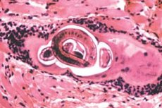
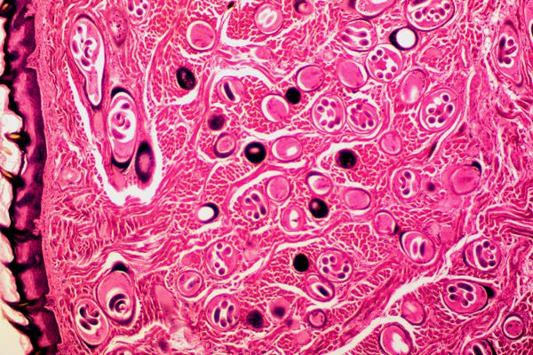
It should be noted that only the larvae that have reached the striated muscles survive, since only skeletal muscle cells can support the parasite. The larva not only hides in such cells from the host's immune system, forming a collagen capsule, but also stimulates the development of blood vessels around the affected cell to obtain the necessary nutrients.
The first larval, infectious stage of Trichinella occurs in the protective cyst; here the anaerobic larva can remain from 15 days to several months or decades, maintaining viability in capsules that calcify and take the form of intramuscular cysts.
 [ 1 ], [ 2 ], [ 3 ], [ 4 ], [ 5 ], [ 6 ], [ 7 ], [ 8 ], [ 9 ]
Life cycle trichinellae
The only ways of infection with Trichinella are food, that is, the parasite enters the human body through the consumption of meat from animals infected with pathogenic larvae enclosed in capsules-cysts. The capsules dissolve in gastric juice, and the larvae freely penetrate the intestinal mucosa, where they develop into adult worms in the course of several molts.
The life cycle of Trichinella occurs in the body of one host (animal or human), and the worm does not need to go outside. Development and colonization of Trichinella spiralis occurs during four larval and one adult stage. The first larval stage occurs in the striated muscles, and in the mucous membrane of the small intestine - three subsequent larval stages (which represent the process of molting) and the stage of the adult worm. The immature small Trichinella feeds on the contents of the mucous cells, damaging them with a stylet, and after 3-4 days is ready to reproduce.
Thus, the life cycle of Trichinella begins with the enteric phase of infection, when a person or animal eats contaminated meat containing first-stage larvae – muscle larvae.
The typical localization of Trichinella is: the masticatory striated muscles of the head; the oculomotor muscles of the orbit and orbital surface of the upper jaw; the diaphragmatic muscles, the skeletal muscles of the shoulder, neck and lumbar region. This may be due to the high level of vascularization of these muscle groups, as well as the significant content of myoglobulin in the sarcoplasm of the membranes surrounding the cells of skeletal muscles.
Pathogenesis
The invasion of the larva through the intestine and its path to muscle tissue causes the pathogenic effect of Trichinella.
Firstly, the movement of the larva, “making” its way to the right place, is accompanied by the inevitable destruction of cell membranes, loss of cytoplasm and damage to organelles, which causes cell death.
Secondly, the migration of newborn larvae with the blood and lymph flow can carry them not only to the tissues of striated muscles, but also to the cells of the liver, kidneys, lungs, myocardium and brain. And the more larvae "wander" around the human body in search of a suitable place in the muscles, the more severe the results of the invasion. This is expressed in general edema, increased excretion of protein in the urine (proteinuria), disruption of calcium metabolism in the body, cardiomyopathy and abnormalities of the central nervous system.
Thus, the pathogenic effect of trichinella can lead not only to parasitic myositis with constant pain, but also to such life-threatening diseases as myocarditis, encephalitis, meningitis, nephritis. Trichinella in children can cause eosinophilic pneumonia or bronchopneumonia, myocarditis, meningoencephalitis. Read more - Trichinellosis in children
[ 12 ], [ 13 ], [ 14 ], [ 15 ], [ 16 ], [ 17 ], [ 18 ], [ 19 ]
Symptoms
Clinical symptoms of trichinellosis largely correlate with the number of larvae that have entered the body, the stage of infection (enteric or muscular), and the state of the human immune system. So the infection may be subclinical.
Initial symptoms of the enteral phase, which may appear 24-48 hours after eating contaminated meat, include general malaise and weakness, fever and chills, hyperhidrosis, diarrhea, nausea and vomiting, abdominal pain, which are caused by the invasion of the intestinal mucosa by larvae and adult worms. These symptoms are non-specific and are characteristic of many intestinal disorders, so in many cases this phase of infection (lasting from two weeks to a month) is diagnosed as food poisoning or intestinal flu.
Symptoms of a Trichonella infestation may slowly worsen as the larvae migrate through the lymphatic system to the muscles. Intestinal symptoms may include cough, headache, swelling of the face and orbital area, conjunctival or retinal hemorrhages, petechiae under the nails, muscle pain, cramps, pruritus, and papular rashes. These symptoms may persist for up to eight weeks.
Severe infection with Trichinella can lead to impaired coordination of hand movements; loss of motor functions (including walking); difficulty swallowing and breathing; weakening of the pulse and decrease in blood pressure; kidney dysfunction; development of inflammatory foci in the lungs, heart, brain; nervous disorders.
Forms
Nematodes of the genus Trichinella infect a wide range of mammals, birds and reptiles. In addition to Trichinella spiralis (parasitic in the body of definitive hosts - domestic pigs and wild boars, other synanthropic and wild carnivores), there are such species of this helminth as: Trichinella nativa, found in polar bears, seals and walruses of the Arctic; Trichinella nelsoni - in African predators and scavengers; Trichinella britovi - in carnivores of Europe, Western Asia and Northwest Africa; Trichinella murelli - in bears, elks and horses in North America.
These species of Trichinella, invading the host's muscle tissue cells, form collagen capsules around the cells with the worm's larvae, which ensure their safe development.
But Trichinella pseudospiralis, a parasite of mammals in temperate climate zones, has a morphological similarity to Trichinella spiralis and belongs to non-encapsulating varieties. Most often, Trichinella pseudospiralis has predatory birds as its main hosts, including migratory ones, which expands the geographical range of the parasite.
Other non-encapsulated Trichinella include Trichinella papuae, a parasite of wild and domestic pigs and saltwater crocodiles in Papua New Guinea and Thailand, and Trichinella zimbabwensis, which infects African reptiles.
Diagnostics
Early clinical diagnosis of Trichinella is quite difficult, since there are no pathognomonic signs. In addition, diagnosis during the first week of infection is complicated by the fact that increased synthesis of the enzymes creatine phosphokinase (CPK) and lactate dehydrogenase (LDH), detected in a blood test, is also observed in other infections.
Serum eosinophilic granulocyte levels also increase, but this is also nonspecific for trichinosis and may indicate other parasitic infections, allergies, or the presence of a malignancy in the patient.
The presence of Trichinella larvae in the body is indicated by antibodies to Trichinella (IgG, IgM and IgE), which can be detected in the patient's blood as early as 12 days after infection - during a serological study of a blood sample using indirect immunofluorescence and latex agglutination methods. More information in the article - Analysis for trichinellosis: antibodies to Trichinella spiralis in the blood
It is possible to detect Trichinella DNA using PCR, but the cost of such testing is too high for most hospital laboratories.
Diagnosis of Trichinella infection also involves a muscle biopsy, for which a tissue sample is taken from the deltoid muscle. However, given the small number of larvae encapsulated in muscle tissue and the 17-24 day incubation period of their development, the result of this study may be false negative.
So, indirect evidence of infection with this parasite can be bilateral periorbital edema, petechial hemorrhages under the nail plates, as well as high temperature in combination with a history of eating undercooked meat.
Treatment
According to experts, treatment of trichinella with anthelmintic drugs is possible only at an early stage of infection, while the parasite is in the small intestine. It is very difficult to expel larvae from muscle tissue with currently available medications.
However, an antihelminthic drug such as Albenzadol (other trade names: Zentel, Gelmadol, Nemozol, Sanoxal) is prescribed - one tablet (400 mg) during meals for 7-10 days. Trichinella is also treated with Mebendazole (Wormin), which is taken 2-4 tablets (0.2-0.4 g) three times a day during the first three days of treatment, and in the following 7 days - three times a day, 0.5 g (5 tablets).
Systemic corticosteroids, in particular prednisolone, are also used simultaneously to prevent exacerbation of inflammatory reactions associated with accelerated elimination of endotoxins (the so-called Jarisch-Herxheimer reaction). And muscle pain in trichinosis is relieved with NSAIDs.
Folk remedies for trichinella
Well-known folk remedies for anthelmintic trichinella will not help if the parasite larvae have already found themselves in muscle tissue. And at the enteral stage of trichinellosis, it is recommended to take decoctions of medicinal plants:
- centaury and elecampane (10 g of each herb per 200 ml of boiling water) - drink several sips throughout the day;
- chamomile flowers, common tansy, lady's mantle and valerian rhizomes - mix a tablespoon of each herb, pour 250 ml of boiling water over a tablespoon of the resulting herbal mixture, boil for 10 minutes, leave under a lid for half an hour; take 100 ml twice a day for 3-5 days.
And to relieve intestinal inflammation during diarrhea, you need to use the rhizome of couch grass, fireweed (narrow-leaved fireweed), knotweed (bird's knotweed) and medicinal speedwell. The mixture of herbs and the decoction from it are prepared as in the previous recipe.
Prevention trichinellae
The main prevention of infection with Trichinella is to eat high-quality meat that has passed sanitary and veterinary inspection, to be especially careful when eating game, and to subject meat to long-term heat treatment. It should be borne in mind that smoking, quick frying (rare steaks), steaming or microwave cooking do not kill Trichinella larvae: meat should be cooked at a temperature of +70-75°C, and it is safest to boil it for a long time.
Increased precautions are required when eating pork. Parasitologists recommend freezing pork at -20°C for 7-10 days (or at -15°C for three weeks) to neutralize this parasite. The thickness of the piece of meat should not exceed 10 cm.
Proper veterinary control of livestock for meat production is extremely important for the prevention of trichinella. In EU countries, according to the decision of the European Commission, since 2005, each batch of meat supplied by producers is tested for trichinella spiralis larvae.