Medical expert of the article

New publications
Treatment of femoral neck fracture
Last reviewed: 29.06.2025

All iLive content is medically reviewed or fact checked to ensure as much factual accuracy as possible.
We have strict sourcing guidelines and only link to reputable media sites, academic research institutions and, whenever possible, medically peer reviewed studies. Note that the numbers in parentheses ([1], [2], etc.) are clickable links to these studies.
If you feel that any of our content is inaccurate, out-of-date, or otherwise questionable, please select it and press Ctrl + Enter.
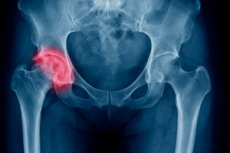
The largest and thickest of all the long tubular bones in our skeleton is the femur. At the top, the bone ends in a rounded articular head or epiphysis, connected to the body of the bone (diaphysis) by the neck. This is the narrowest place of the femur, and a fracture of this localization is a fairly common injury, especially in the elderly, which is due to the age-related decrease in bone strength. Treatment of femoral neck fracture is most often surgical and is accompanied by long-term rehabilitation - on average, this period takes six months from the moment of surgery. In cases where the nature of the injury allows avoiding surgical intervention and the patient's age suggests that the femoral neck will heal on its own, conservative therapy may be used.
However, treatment without surgery is associated with prolonged forced immobility of the patient, which leads to the development of complications. In the elderly, these include pressure sores, psychoemotional disorders, deep vein thrombosis and hypostatic pneumonia, which may cause the patient's death. In addition, there is a high risk of bone non-union in age patients. Therefore, surgical treatment of femoral neck fracture specifically in older victims who were walking before the injury is used for vital indications.
In young and middle-aged patients, prolonged bed rest is also difficult to tolerate, and conservative treatment often does not lead to the desired result and is just a postponement of surgery. Moreover, in young patients, fractures are more often complex, resulting from significant traumatic effects, such as falls from a great height or automobile accidents. Therefore, surgical treatment is the method of choice in most cases of femoral neck fractures in patients of any age.
Timely medical attention (immediately after a fracture) is the key to successful treatment. In complex fractures of the femoral neck, the person cannot walk, has severe pain up to shock, the injury in such cases is usually caused by a high-energy impact, which makes it necessary to seek help immediately.
However, in elderly patients with sparse bone tissue, a fracture can occur even from an unfortunate rollover in bed, a sudden bend, or a minor impact, such as on the edge of a table. Symptomatology in such cases is weak, and the patient does not assume the presence of a fracture. He continues to walk, limping, treated for radiculitis or osteochondrosis folk remedies, and during this time the condition of the femoral articulation worsens - there is a displacement, finally disrupted blood supply and develops aseptic necrosis of the articular head. Therefore, in the case of a sudden appearance of new sensations in the area of the hip joint, it is better to show concern and immediately undergo examination.
The following symptoms should alert: not too strong, but constant pain in the groin area, which increases when trying to walk faster, climb stairs or step on the heel; crunching and difficulty in turning the lower body in the supine position; in the same position one can notice a shortening of the length of the affected leg and a noticeable turning of the foot with the toe outward (the outer side of the foot touches the plane of the bed). Typical is the symptom of "stuck" heel, when the patient can not tear it from the horizontal surface in the supine position, but is able to bend and straighten the knee. Additionally, you can independently with the help of loved ones to conduct verification tests: ask someone to press or tap on the heel - such actions are usually responded by pain in the groin or pelvic area. It also occurs when palpating the hip joint on the affected side. Should be alerted to the sudden appearance of a hematoma - when a fracture is damaged vessels located in the depths, so the blood to the surface of the skin does not penetrate immediately, but after some time, and the appearance of a bruise is not directly preceded by a blow. These signs - a reason for immediate examination. Time is working against you. [1]
When choosing treatment methods for a femoral neck fracture, the doctor takes into account many factors: the type and localization of bone damage, the patient's age, his or her state of health, and the degree of neglect of the problem. Only after a comprehensive examination and a complete collection of anamnesis is the question of the preferred treatment tactics decided.
Classification of femoral neck fractures is performed according to several criteria reflecting the clinical nature of the injury. According to the location of the neck bone fracture line relative to the epiphysis, they are subdivided into basicervical (in the lower part of the neck, at its base, base), transcervical (approximately in the middle), subcapital (above, under the head itself). This characteristic indicates the degree of risk of aseptic necrosis - the higher the fracture line, the more disturbed the epiphyseal blood supply and less likely to fuse the bone independently, i.e. Urgent surgery is more relevant.
The chances of recovery also depend on the angle of the fracture line to the vertical axis (Powels classification). The least favorable location is when this angle is less than 30° (fracture complexity grade I). The femoral neck is considered more viable when the angle is between 30° and 50° (Grade II). Close to horizontal location of the fracture line is the most prognostically favorable (III degree, angle of more than 50°).
Subcapital, the most dangerous fractures of the femoral neck, are in turn classified according to Garden into four types. The most complex is the fourth, complete (completed) fracture with displacement of the fragments, in which case they are completely separated; the third type includes completed fractures with partial retention of the fragments and partial displacement; the second type includes complete fractures without displacement; the first type includes incomplete fractures, so-called bone cracks, which have the shape of a green twig. The latter are well amenable to conservative treatment in timely treatment, but in neglected cases, if the patient tolerates discomfort and continues to walk, pass into a complete fracture.
In addition, according to the type of displacement of the epiphysis fragments, there are varus (downward and inward), valgus (upward and outward), and embedded, in which (a neck fragment falls inside another). The latter can be confused on X-ray with an incomplete fracture. Computed tomography, for example, is used to differentiate between the two. A femoral neck fracture is complete, but it has a favorable prognosis and can be cured conservatively with timely treatment.
Treatment of pareloma of the femoral neck with surgery
Surgical treatment is the method of choice for any type of fracture. It is the most effective method. The injury is severe, bone fusion in a patient of any age, even with a favorable prognosis is still questionable. Therefore, if the patient was walking before the fracture and his health condition allows him to undergo a major surgery, and if osteosynthesis is used - two, since the metal structures are removed after 1.5-2 years, surgical treatment is preferable.
There are two main techniques used in the surgical treatment of a fracture - osteosynthesis and endoprosthesis. The choice between the two is less about the type of fracture and more about the patient's age and level of physical activity prior to injury. In younger and healthier patients, on average up to the age of 60, osteosynthesis is used to preserve all the natural components of the hip joint. In the elderly and senile age, the blood supply to the bone tissue is already impaired as well as the ability to restore its integrity, so endoprosthesis is considered the preferred operation. It is for age patients that such an operation is the only chance to restore motor activity. [2]
Contraindications to surgery include:
- Poor somatic or mental health, exhaustion, i.e. There is a high probability that the patient will not tolerate the operation;
- Internal bleeding, clotting problems;
- Infection of the surgical area;
- Venous insufficiency of the affected limb;
- Systemic bone disease;
- Severe chronic and acute pathologies (diabetes mellitus, recent heart attack or stroke, severe musculoskeletal disorders, etc.).
If the patient was not walking before the fracture, surgery is not even considered as a treatment option. If the patient is overweight, surgery may also be an obstacle. [3]
Osteosynthesis
This technique consists in restoring the integrity of the hip joint using various fixation structures. The bone fragments are placed in the correct position and firmly fixed with fixators (pins, screws, plates) made of inert materials until complete fusion.
In the absence of fragments and displacement, osteosynthesis is performed in a closed method - through a small incision without opening the joint capsule under the control of a radiological apparatus and an electron-optical converter, or in complex fractures requiring full access - open. During surgery, the patient is under anesthesia, general or spinal.
Currently, osteosynthesis is rarely used. This is primarily due to the fact that most patients with this injury are elderly. Osteosynthesis is suitable for younger patients, because the hip prosthesis has a shelf life, after which it must be replaced. And this is a new operation and, the younger the patient, the more they will have to do in the future. Also, if the fracture of the femoral neck occurred in childhood or adolescence, they try to save the natural joint, which will still grow. [4]
Indications for osteosynthesis surgery are: femoral neck fragment fracture, the presence of displacements, fracture of the I degree of complexity, a combination of fracture and dislocation, ineffectiveness of conservative therapy or previous surgical intervention, and also taken into account:
- Tissue viability of the femoral head;
- The age of the patient (on average up to 60 years old);
- His activity and mobility prior to the injury;
- Inability to fit a prosthesis.
The osteosynthesis method is used mainly for the treatment of embedded, transcervical and basal fractures, but also for subcapital fractures in young patients.
Bone fragments are joined using two methods: intraosseous (intramedullary) and periosteal (extramedullary). In complex fractures, these two methods are combined. The fixation structures are placed in such a way that a firm contact of the fractures in an anatomically correct position is ensured. Fasteners are selected according to the architectonics of the bones of the hip joint, they are rigid or semi-elastic, making it possible to fix multiple small fragments. Modern fasteners are made of inert, biologically compatible alloys based on steel or titanium.
Intramedullary (immersion) osteosynthesis is more commonly used, where pins are inserted through the medullary canals of the distal and proximal fragments to connect them. The ends of the pins usually have screw holes or are bent in a certain way to create a stable immobilized structure. Sometimes the canal is drilled out to insert the pin.
After bone fusion, all fixation devices are removed. The operation to remove them is usually not associated with complications.
The extramedullary (periosteal) method consists of placing rings on the outer surface of the bone, a plate fixed with screws, and suturing the fragments with serclage sutures.
Intramedullary fixators as well as periosteal sutures and rings usually require additional fixation measures such as limb plastering. Extramedullary plates provide stability by themselves. [5]
Osteosynthesis surgery should be performed as soon as possible, preferably within the first day after the fracture. Examination of the patient is done according to an accelerated program. It includes laboratory and instrumental studies. The operation itself is performed under general or spinal anesthesia. During surgical intervention, surgical X-ray control is performed in the anteroposterior and axial projection of the joint.
Immediately after surgery, the patient is prescribed a course of antibacterial drugs, since a deep invasive intervention was performed. This tactic helps to prevent infectious complications. Also prescribed are painkillers, vitamins, drugs with calcium and to activate blood circulation. Depending on the specific situation, anticoagulants, immunomodulators, nonsteroidal anti-inflammatory drugs, glucocorticosteroids may be prescribed. [6]
The patient is activated from the second day after the operation - he starts walking with the help of crutches.
In addition to infection, the following complications can occur after osteosynthesis surgery:
- Stability of fixation, separation of the fragments;
- Intra-articular hematoma;
- Blood supply problems and, as a consequence, the femoral neck and femoral head never fuse, the latter is destroyed (aseptic necrosis);
- The formation of a false joint;
- Osteomyelitis;
- Arthritis/arthritis of the hip joint;
- Deep vein thrombosis in the affected leg;
- Pulmonary embolism;
- Hypostatic pneumonia.
Minimizing the likelihood of complications is facilitated by a clear implementation of the rehabilitation program. [7]
Endoprosthetics
Nowadays, hip replacement with a prosthesis is recommended more often for preserved elderly and elderly patients with femoral neck fracture. This operation gives the injured person the ability to move fully. An indication for surgery is the patient's advanced age, which suggests that the fracture will not heal due to impaired blood supply. Treatment of femoral neck fracture with displacement in age patients by endoprosthesis is vital and allows to prevent disability, especially if there is a marked displacement of fragments and a large number of fragments, aseptic necrosis, degenerative-dystrophic changes of the joint, its inflammation, etc. In addition, recovery after endoprosthesis takes a shorter period than after osteosynthesis.
Contraindications, common for operations to restore the mobility of the TBS (hip joint), when it is necessary to replace the "native" joint with an implant are considered individually.
The choice of prosthesis depends on many factors. The main one is the mobility of the patient before the injury and the condition of the bones. For those who left the house and moved freely without special restrictions, it is recommended to install bipolar (total) endoprostheses, which involve replacing not only the head and neck of the femur, but also the acetabulum. In elderly patients (on average over 75 years of age), who before the injury had limited mobility, either in an apartment or not far from home, unipolar (subtotal) endoprostheses are recommended, replacing only the femoral head and neck, which is placed in the natural acetabulum. [8]
The artificial implant completely repeats the shape and dimensions of the native joint and is made of durable inert material: the cup (acetabulum) is usually metal with a ceramic or polymer insert; the epiphysis (head) is made of a metal alloy with a polymer coating; the neck, which transitions into the stem, as the most loaded part, is also made exclusively of durable alloys.
The following methods of endoprosthesis fixation are used:
- Cementless - implant with a porous coating, tightly fitted into place, with subsequent bone sprouting into it;
- Cemented - fixed in place with a special cement made of a polymer material;
- Combined - the bone head is cementless and the stem is cemented or in young patients with a bipolar prosthesis, the cup replacing the acetabulum is additionally secured with screws.
Older patients with osteoporosis are usually cemented with a denture.
Briefly, the process of surgery is done in stages. The patient is put under anesthesia. After providing surgical access to the joint, the parts to be replaced are removed, the prosthesis is installed and fixed, a drainage tube for the outflow of fluid is installed, then the layers of muscle and skin are sutured, starting from the deepest and a soft dressing is applied. On average, endoprosthetic surgery lasts two to three hours. [9]
Immediately after surgery, the patient is prescribed antibiotics and painkillers, other medications and procedures - according to symptoms.
In addition to infection and healing problems, complications of hip arthroplasty include rare cases such as implant rejection and periprosthetic fracture of the femur, which occurs below the level where the prosthesis is inserted and is caused by insertion errors. People with sparse bone tissue (osteoporosis) are more likely to suffer from the second complication. [10]
The service life of a quality Tibial Prosthesis is usually more than 10-12 years, but it still has to be replaced at some point. The moving parts of the prosthesis are subjected to wear and tear caused by friction. This is the main disadvantage of endoprosthetics.
In other respects, this operation has a number of advantages over osteosynthesis: faster rehabilitation (on average it takes 2-3 months), in general - fewer complications. [11]
Treatment of femoral neck fracture without surgery (conservative treatment)
Surgical treatment is the method of choice for any femoral neck fracture for patients of any age. After surgical intervention, a person definitely recovers faster, gets back on his feet and starts walking.
Theoretically, treatment of femoral neck fracture without displacement can be carried out by conservative methods, and it was done in the past, but treatment without surgery has not justified itself. It is not easy for a young healthy person, and for the elderly such consequences as bedsores, thromboembolism, hypostatic pneumonia, depression can lead to premature death.
Nevertheless, a certain contingent of patients is contraindicated for health reasons. These are people with severe systemic pathologies, who will not tolerate anesthesia. It makes no sense to perform surgery on patients who did not walk before the fracture. Sometimes even young people with a fracture of the femoral neck for various reasons refuse surgery or have contraindications to it.
Conservative treatment can be effective if the fracture is incomplete or the fracture line is located at the base of the neck and is nearly horizontal, there is no displacement, the patient is young enough, and there are no problems with the blood supply to the proximal fragment.
Non-surgical treatment of a non-dislocated femoral neck fracture without displacement may also be successful.
Timely therapy is important, which consists of skeletal traction of the injured limb and immobilization by applying a plaster cast. The course of therapy also includes taking medications prescribed by the doctor, massage, breathing and therapeutic exercises, and the use of apparatus physical methods.
Treatment of medial femoral neck fracture (i.e., intra-articular) without endoprosthesis rarely has a favorable prognosis, much less conservative treatment. When the fracture line is located in the middle and upper part of the femoral neck, there is a high probability of complete cessation of blood supply to the femoral head and its subsequent necrosis. Even osteosynthesis is rarely recommended for this type of fracture.
As a result of prolonged conservative treatment, even in young patients, true fusion does not occur in most cases. A connective tissue callus forms in the fracture area, which holds the fragments together. Even after osteosynthesis, the bones often do not fuse, but they are held together by a stronger structure. The function of the joint therefore remains impaired to varying degrees.
However, if the patient has categorical contraindications to surgery (mentioned above), the type of fracture does not matter. In any case, the patient must be prescribed and treated, the main purpose of which is to prevent and eliminate complications associated with prolonged immobility: pressure sores, muscle atrophy, thromboembolism, hypostatic pneumonia. The hospital stay in the treatment of a non-surgical femoral neck fracture is usually at least three months.
If the age and bone condition of the injured person gives hope that the bones will fuse, the following treatment tactics are used. First of all, skeletal traction is performed on the injured limb. The procedure has different goals depending on the type of fracture: in the case of a splinter fracture - repositioning of the fragments, in the case of an embedded fracture - allows the bone to fall into place and prevents shortening of the leg. Traction can be the main method of treatment or additional before immobilization of the limb, respectively, and the duration of this stage can be different - from ten days to two or more months.
The main method of treatment is used for femoral neck fractures: traction is performed immediately after the injury. The leg is anesthetized and placed in a special Belair splint, to which a weight of approximately three kilograms is attached. The patient's leg is elevated and moved away from the center line of the body. The patient's head is also raised. After about two months, the traction is removed. The patient is allowed to move with crutches without leaning on the affected leg. After another two months, under the supervision of a doctor, the patient begins to gently use the affected limb when walking. The entire treatment period takes about 6-8 months.
Immobilization is used for basocervical fractures. Skeletal traction is applied to the affected limb when there is a dislocation of the fragments for ten days or two weeks (in the case of a non-displaced fracture, fixation is performed immediately). The hip joint is then fixed with a plaster cast for a period of three months or more: a corset is applied to the abdominal area and a plaster cast for the broken leg is attached (coxit bandage). It is fixed in a position slightly to the side. Sometimes the plaster cast will have to be worn for more than six months. After the cast is removed, the patient can walk on crutches without leaning on the leg. A derotation boot is used to fix the sore leg and reduce soreness. When the X-ray shows that the bone has fused, you can begin to gradually load it.
Such methods of bone fusion are rarely used, as they are associated with prolonged immobility and the many complications it causes. To prevent them, from the very first days, immobilized patients are prescribed breathing exercises, physical therapy, and massage. It is necessary to work as a sick leg, as well as a healthy one. It is recommended to actively work the feet and toes, tense the muscles of the thigh and ankle, perform bends and turns of the head and torso, squatting in bed. Early activation of the patient is facilitated by a Balkan frame for the treatment of fracture of the femoral neck, which is usually equipped with a bed in the orthopedic department. It is a device that allows the patient to pull himself up on his arms and squat independently in bed, as well as to do some exercises of therapeutic gymnastics.
Weak elderly patients who are contraindicated from surgery for health reasons are treated without such painful procedures as skeletal traction, immobilization and repositioning of fragments. So-called functional treatment is used. The patient is hospitalized, the main mode - bed rest. In the supine position, he is put a roller under the knee to keep it in an elevated position, which limits the rotation of the leg. Painkillers are prescribed.
Literally from the first days, the patient's early activation tactics are carried out: he or she is seated in bed using a Balkan frame, turned on his or her side, and taught to walk on crutches or with a walker. The bones in these patients do not fuse, the limb is shortened, external rotation remains, and they have to walk on crutches for the rest of their lives. However, because they remain active, they do not develop life-threatening complications.
Rehabilitation
The recovery period begins immediately after surgery, and in conservative treatment it is difficult to distinguish it from treatment at all. At present, early activation of patients is preferred, as passive recumbent lifestyle leads to muscle atrophy and development of severe complications.
Rehabilitation measures include drug rehabilitation therapy, therapeutic exercises, massage, apparatus physiotherapy (electro- and magnetotherapy directly through the plaster cast), a certain diet, hygienic procedures, prevention of pressure sores and congestion.
Both in conservative treatment and after surgery, the patient is prescribed vitamin and mineral complexes to accelerate fusion, formation of bone and connective tissue callus, restoration of impaired blood supply and prevention of degenerative-dystrophic joint changes. There are no specific drugs for TBS, complexes are selected individually, but their mandatory elements are calcium, vitamin D, chondroitin and glucosamine.
Fractures may be accompanied by pain. In this case, non-narcotic analgesics from the group of NSAIDs are prescribed, which also relieve swelling, thin the blood and control inflammation. Patients prone to thrombosis are recommended to take anticoagulants, those suffering from edema - anti-edema agents.
Immunostimulants may be prescribed for open fractures and elderly people with reduced immunity, homeopathy, phytotherapy and bioactive food supplements are also used to accelerate healing.
The complex of drugs should be prescribed by a doctor individually. The patient should follow the recommendations received, do not be amateurish, follow the rules of intake, since the interaction of some drugs can either weaken their effects or lead to undesirable results.
Therapeutic massage is prescribed immediately after radical measures (surgery, skeletal traction, immobilization), and continues even after the fixation bandage is removed. In the hospital, it is performed by a qualified specialist. Massage the patient not only the injured limb and the lumbar area above the cast, but also the chest (prevention of congestive pneumonia), healthy leg (prevention of atrophic process), feet and shins. General massage improves blood circulation, which helps to accelerate the healing of the injury.
Therapeutic exercises. It is also carried out initially under the supervision of a physiotherapist, instructor or attending orthopaedic doctor. Exercises for patients who are not too mobile are selected in such a way that almost all muscle groups are involved. These are head turns in different directions, exercises with weighted hands, movements of feet and toes (stretching, compression, rotation), a healthy leg can simulate riding a bicycle, bending and extending it, tense the muscles of the limbs, gluteal, abdominal muscles. It has been proven that even mentally performing exercises causes blood flow to the involved organs and trains them.
Breathing exercises are also performed: the usual cheerful singing, inflating balloons, exhaling air into a glass of water through a tube, etc. Breathing exercises prevent congestion in the lungs and the development of hypostatic pneumonia. Loads when performing physical exercises should be feasible, the patient should not overwork, but passivity is not welcome.
The patient's diet should contain an optimal amount of proteins, fats and carbohydrates, and vitamins, in particular calcium (bananas, fermented milk products) and vitamin D (fish, eggs, cod liver), contain enough fiber (raw fruits and vegetables, whole-grain bread) to activate intestinal peristalsis. Feed the patient preferably in small portions 5-6 times a day. Give to drink plenty of fluids. Prefer to give preference to dishes stewed, steamed or cooked in the oven. Limit spicy, fatty, fried, exclude alcohol, sweet carbonated drinks. In short, follow the general rules of healthy eating.
To prevent pressure sores, special orthopedic bedding is used, and hygiene of the body, clothing and bedding is observed. The skin in places of pressure and friction is treated with special preparations or just camphor alcohol.
Carefully observed hygiene of the oral cavity, intimate areas, the whole body - the patient is brushed, washed, washed, assisted in brushing teeth, serve a vessel or change diapers.
After the patient is discharged home, all rehabilitation activities continue.
Recovery time depends on many factors: the type of fracture, the timing of first aid, the chosen treatment method, the age of the injured person, the condition of his/her bone tissue and its ability to regenerate, the general medical status, the desire to recover and active conscious participation in the rehabilitation process.
Patients who have undergone endoprosthetic replacement are the fastest to recover from a femoral neck fracture and generally have fewer complications. Only very mild fractures can be fully recovered with conservative methods, in most cases there is no full recovery. Osteosynthesis occupies an intermediate position between the two methods. On average, it takes six months from the time of fracture to full recovery, but in patients with chronic diseases it may take a year or a year and a half. The risk of complications increases in diabetics, cancer patients, people with thyroid problems, smokers and drinkers, poor diet, osteoporosis and other degenerative bone and joint processes. This is by no means a complete list of risks. Much depends on the mood of the patient: sometimes a patient of very old age fully recovers, and younger, but passive, pessimistic attitude and walks with a stick, limping. In general, though, younger patients take less time to recover than older patients.
A fracture of the femoral neck is not a verdict. Modern medicine and the desire to recover, as well as the help of people close to you can work wonders. Prevention of TBS injuries, especially in old age, is also important. Especially if there has already been a hip injury. Such people need to be careful when walking on stairs - stick to the railing, in winter use anti-slip devices for shoes, try not to leave the house in the ice. Also will help to avoid injury weight within normal limits and moderate physical activity, balanced diet, absence of bad habits, taking vitamin and mineral supplements enriched with calcium and vitamin D, drugs that prevent loss of consciousness, because in old age many people suffer from coronary heart disease, cerebrovascular disease, pressure fluctuations.
Literature used
Vygovskaya O.N. Principles of care for femoral neck fracture, Novosibirsk, 2016
Dmitry Naidenov: 99 Tips for Hip Neck Fracture, Nevsky Prospect, 2011
Sergei Ivannikov, Nikolay Sideshow, Yusef Gamdi. Fractures of the femoral neck, 2005