Medical expert of the article
New publications
Transvaginal cervicometry of the cervix: how it is performed and how often it is done
Last reviewed: 03.07.2025

All iLive content is medically reviewed or fact checked to ensure as much factual accuracy as possible.
We have strict sourcing guidelines and only link to reputable media sites, academic research institutions and, whenever possible, medically peer reviewed studies. Note that the numbers in parentheses ([1], [2], etc.) are clickable links to these studies.
If you feel that any of our content is inaccurate, out-of-date, or otherwise questionable, please select it and press Ctrl + Enter.

Cervicometry refers to a procedure designed to determine the length of the cervix. A special ultrasound machine is used for this. This data must be known in order to further predict the course of pregnancy and understand how the fetus is held inside the uterus. If the indicators are normal, then there is no reason to worry. If the length is shorter than required, there is a risk of developing serious pathologies, in particular, premature birth. Cervicometry makes it possible to promptly identify many pathologies that occur during pregnancy and prevent a number of dangerous pathologies. Knowing the results, you can take the necessary measures in a timely manner and prescribe the required treatment, which will prevent the risk.
What is cervicometry during pregnancy?
This is one of many diagnostic procedures used to determine the risk of developing possible pathologies and complications. It can be performed in two ways - internally and externally. Each method has its advantages and disadvantages, so the choice is always up to the doctor. Most specialists are inclined to believe that transvaginal cervicometry should be used to obtain more accurate results.
For external examination, the length of the cervix is recorded using a traditional ultrasound device. It is measured through the peritoneum. With a full bladder, it becomes possible to more accurately palpate the uterus and cervix.
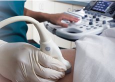
There is also a method that is more accurate – the transvaginal method. It is performed with an empty bladder to ensure greater accuracy of the results. When urine accumulates, it is not possible to fully view the entire picture and take measurements. The study is based on the use of a special transvaginal sensor, which is inserted directly into the vagina. The cervix is examined, important indicators are measured. For the doctor, it is absolutely unimportant which method is used to take measurements, the result itself is important.
A routine examination includes an ultrasound scan, during which measurements are taken (18-22 weeks). This is usually enough, but if there is a risk of developing ICI, previous miscarriage and premature birth, miscarriages, then it is necessary to conduct a transvaginal examination. If the indicators do not correspond to the norm, it is necessary to take urgent measures, otherwise there is a risk of termination.
Is cervicometry harmful?
The manipulation is harmless to the fetus and mother, absolutely painless. Ultrasound exposure is minimized for safety purposes. This was achieved by reducing the power of the waves and shortening the duration of the procedure. The woman should not worry at all, since all the nuances in modern equipment have long been taken into account.
Specialists use the device in a special energy mode, in which the impact is limited, which leads to a limitation of the acoustic power, as a result of which no additional influence occurs.
Indications for the procedure
The procedure is performed, first of all, when premature birth occurs, or they were observed earlier, with an increased probability of miscarriages. It is performed in case of abnormal development of the uterus, to diagnose ICI. The procedure is mandatory for those who are carrying several babies, or twins. For insurance, it is performed if the woman has undergone surgical interventions of any nature or direction: whether they are preventive, for the purpose of treatment or diagnosis. Regular measurements are carried out to monitor the condition of scars, uterine sutures.
 [ 1 ]
[ 1 ]
Preparation
In the process of preparation for cervicometry, no measures are required. It is only necessary to empty the bladder if the study is performed transvaginally, and to maintain its fullness during external examination. This is necessary in order to obtain the most accurate results. No other preparation measures are required, since everything necessary will be done by the doctor conducting the study. You do not even need to worry about the results: the specialist will make a conclusion and provide it to the obstetrician-supervisor.
Technique cervicometry
First, the patient must completely empty her bowels, then lie down in the lithotomy position (traditionally on a gynecological chair). The essence of the procedure is the introduction of a special sensor into the vaginal environment, which allows for an examination with the necessary measurements, records the result and displays the image on the computer.
Each measurement lasts on average 2-3 minutes. The size of the cervix may change by approximately 1% depending on uterine contractions. If the values differ, the shortest option is taken into account. In the second trimester, the fetus is mobile, and the values vary (this depends on the position of the fetus). The results are most variable in the area of the uterine floor and in the transverse position of the fetus.
There is another method of assessing the size of the uterus, in which measurements are taken transabdominally. This is an external method. But it can be called a visual assessment rather than cervicometry. The indicators obtained with this method of measurement are unreliable, they differ significantly from those in reality. The error is 0.5 cm or more, which is significant.
Cervicometry of the cervix
It is necessary to know the size of the uterus in order to ensure a successful delivery. The course of pregnancy and the ability to bear a child depend primarily on the size. If the cervix is shortened, it may not withstand the pressure of the fetus and begin to open prematurely. This usually ends in miscarriage, spontaneous abortions, premature birth.
The length can also determine the approach of labor. The closer to labor, the shorter the birth canal becomes, and the smaller the size of the cervix. This is a natural and normal process. At each stage of gestation, the indicators are different.
Measurements are taken externally or internally. Only the internal method is accurate. Immediately before labor, the size of the cervix reaches 1 cm, and it gradually begins to open. Throughout the pregnancy, the cervix is covered by a mucous plug, which comes off after the opening process begins. This is normal before labor, but this process can begin at any time, which is not normal and is due to the insufficient size of the cervix. It is necessary to monitor the size throughout the pregnancy in order to be able to take the necessary measures in a timely manner. Additionally, with the help of cervicometry, it is possible to determine the length of all those organs that are related to the labor process. It is also possible to determine the beginning of the opening, if it occurs prematurely. There are often cases when the length of the cervix is normal, but its opening is already happening. In this case, it is possible to take timely measures that will allow you to save the child.
Transvaginal cervicometry
The internal method allows obtaining significant information about the length of the cervical canal. A transvaginal sensor is used for this. The bladder must be empty. Then the patient lies down on the chair, the sensor is inserted into the vaginal cavity. The image is displayed on the monitor. The manipulation is carried out several times, usually three times, which eliminates the possibility of error. The average duration of one measurement is several minutes. The smallest indicator is taken into account. If the result is questionable, light pressure is applied to the lower abdomen for 15 seconds, then the measurements are repeated.
Some specialists resort to the use of electronic digital calipers, which make it possible to measure the size of the pharynx. It is also necessary to take into account individual characteristics. Thus, the norm for primiparous and multiparous women differ significantly.
Cervicometry in dynamics
Sometimes it is necessary to take measurements dynamically. This is necessary if the cervix is sutured and requires monitoring, if the cervical canal is dilated or fetal membranes penetrate into it. The indicators must be taken into account if there were previous premature births or surgical interventions. Insurance is provided for primiparous women or if there is insufficient information. Dynamic indicators are measured once every 14 days.
How often is cervicometry performed?
If there is a need for regular measurements, they are performed at intervals of 14 days. This situation applies to 15% of pregnant women. Usually, the indicators are measured dynamically starting from the 15th week. In the absence of pathologies, the procedure is performed once, at a period of 20-24 weeks.
Normal performance
There are no uniform norm values. They vary significantly and depend on the period, position of the fetus, and whether the pregnancy is the first or repeated. There are many additional factors that also affect the norm values. If measurements are taken at 20 weeks, the norm values will be 40 mm, at 34 weeks, they will decrease to 34 mm.
 [ 6 ]
[ 6 ]
Reviews
Many women leave positive reviews. Firstly, they note that the procedure is painless. Secondly, a big plus is that the results can be obtained quite quickly and you don’t have to torment yourself with fears. Or, on the contrary, if a pathology is detected, you can take the necessary measures in a timely manner. No effect on the future child was found.
There are reviews when this procedure was done to non-pregnant women. This is also possible for diagnosis and treatment of many diseases. The fact is that cervicometry is performed not only to take measurements. You can get an image of the cavity, look at the walls, tissues, conduct an analysis of cervical fluid (daily measurements), which is of great diagnostic importance.

