Medical expert of the article

New publications
Toxoplasmosis chorioretinitis.
Last reviewed: 04.07.2025

All iLive content is medically reviewed or fact checked to ensure as much factual accuracy as possible.
We have strict sourcing guidelines and only link to reputable media sites, academic research institutions and, whenever possible, medically peer reviewed studies. Note that the numbers in parentheses ([1], [2], etc.) are clickable links to these studies.
If you feel that any of our content is inaccurate, out-of-date, or otherwise questionable, please select it and press Ctrl + Enter.
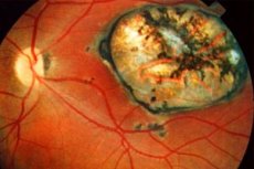
Toxoplasmic chorioretinitis is most often associated with intrauterine infection. Clinical manifestations of eye damage are not always detected at birth and in early childhood.
Congenital toxoplasmosis, like other congenital infections, is characterized by a combination of eye damage with other systemic disorders, most often with CNS damage. Infected newborns may experience fever, lymphadenopathy, encephalitis, hepatosplenomegaly, pneumonia, and intracranial calcifications.
Pathogens
Symptoms toxoplasmosis chorioretinitis.
Symptoms of toxoplasmosis depend on the age and immune status of the patient, as well as on the activity of the eye infection. Toxoplasmosis manifests itself as chorioretinitis. Inactive toxoplasmosis reveals old large atrophic or cicatricial chorioretinal foci with hypertrophy of the pigment epithelium, often solitary, located in the area of the posterior pole of the eye. The appearance of a zone of active inflammation in the form of white foci is observed in any area of the fundus, as a rule, at the edge of old changes. In the acute period of inflammation, the foci have unclear boundaries, their size varies and can be equal to several diameters of the optic nerve disc. With large lesions, their protrusion into the vitreous body is possible. The vessels in the lesion can close. With active inflammation, exudative retinal detachment and secondary choroidal neovascularization with subretinal hemorrhage are possible, visible during ophthalmoscopy as a thickening of grayish-yellowish tissue at the level of the pigment epithelium.
Changes in the vitreous body, infiltration of its layers by cellular suspension and formation of membranes are observed when the process spreads to the inner layers of the retina and the destruction of the hyaloid membrane. In this case, damage to the optic nerve and cystic edema of the macular are noted.
Diagnostics toxoplasmosis chorioretinitis.
Diagnosis is based on the identification of characteristic signs of congenital toxoplasmosis and the typical localization of large single foci in the area of the posterior pole with the formation of new zones of inflammation along the edge of old scars.
Serological testing includes the determination of specific antibodies in toxoplasma using the complement fixation reaction and fluorescent antibodies. The most informative and widely used in recent years is the study with the enzyme immunoassay, which allows the detection of antibodies of different classes.
 [
[ What do need to examine?
How to examine?
What tests are needed?
Who to contact?
Treatment toxoplasmosis chorioretinitis.
Not all forms of toxoplasmosis require treatment. Small peripheral lesions may be asymptomatic and self-heal within 3 weeks to 6 months. In the case of severe symptoms of inflammation in the posterior pole of the eye, as well as in the case of reactivation of the process, treatment should be aimed at destroying microorganisms. Local non-specific anti-inflammatory therapy (corticosteroids) in combination with systemic use of specific agents is indicated.
The drugs most widely used in the treatment of toxoplasmosis include fonsidor, pyrimethamine, daraprim, tindurine, chloridine and sulfadiazine. Treatment is carried out with sulfanilamide drugs in combination with folic acid under the control of blood composition due to the possibility of developing leukopenia and thrombocytopenia. It is possible to use pyrimethamine and sulfadiazine in combination with corticosteroids under the conjunctiva. Clindamycin and dalacin as protein synthesis blockers in the treatment of toxoplasmosis are also used in combination with the drugs described above.