Medical expert of the article

New publications
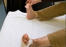
Diabetic foot symptoms
Last reviewed: 06.07.2025

All iLive content is medically reviewed or fact checked to ensure as much factual accuracy as possible.
We have strict sourcing guidelines and only link to reputable media sites, academic research institutions and, whenever possible, medically peer reviewed studies. Note that the numbers in parentheses ([1], [2], etc.) are clickable links to these studies.
If you feel that any of our content is inaccurate, out-of-date, or otherwise questionable, please select it and press Ctrl + Enter.
Clinical features of neuropathic and ischemic forms of diabetic foot syndrome are presented in the table.
In order to decide on the need for antibiotic therapy, timely recognition of systemic and local signs of wound infection is of great importance.
Systemic signs of wound infection:
- fever;
- intoxication;
- leukocytosis.
Local signs of wound infection
- for acute wounds:
- hyperemia;
- edema;
- pain,
- local hyperthermia;
- purulent exudate;
- for chronic wounds:
- pain in the wound area and surrounding tissues;
- bleeding of granulation tissue;
- unpleasant odor;
- increase in wound size;
- profuse exudation;
- slow healing;
- atypical color of granulation tissue;
- formation of cavities at the bottom of the wound.
Clinical signs of osteoarthropathy:
- Acute stage:
- hyperemia;
- hyperthermia (difference of more than 2° C during thermometry);
- swelling;
- pain (in approximately 50% of patients);
- the changes are asymmetrical, usually one-sided;
- X-rays can reveal fractures, dislocations of small bones and joints of the foot;
- Chronic stage:
- deformation of the foot up to the collapse of the arch of the foot;
- changes in foot radiograph;
- Ulcer formation is possible in areas of excess pressure.
Clinical features of neuropathic and ischemic forms of diabetic foot syndrome.
| Sign | Neuropathic form | Ischemic form |
| Middle age | Up to 40 years | Over 55 years old |
| Duration of diabetes | More than 5 years | 1-3 years |
| Other late complications of diabetes mellitus | They meet often | May not be expressed |
| Cardiovascular diseases | Microangiopathy may not be present | Arterial hypertension, hypercholesterolemia, coronary heart disease |
| Bad habits | More frequent alcohol abuse | Smoking more often |
| History of foot ulcers | Often | Rarely |
| Condition of ulcers | Usually painless. Hyperkeratosis of surrounding tissue. | Painful dry necrosis in the form of a scab. Hyperkeratosis of the surrounding tissue is not typical (but fibrin deposition in the form of a "halo" is possible). The skin around the ulcer is thinned, hyperemic (even in the absence of infection) |
| Localization of ulcers | In areas of increased pressure (often caused by foot deformation) - most often on the sole, in the spaces between the toes | In the "acral" zones of the foot - more often on the toes, heels ("acral" necrosis) |
| Leg condition | The skin is pink, warm, dry. Pulsation in the arteries is preserved, the veins are full-blooded. At night, severe pain and paresthesia (restless legs syndrome) may bother. | Skin pale or cyanotic, cold, moist. Pulsation in arteries is reduced or absent. Intermittent claudication or pain at rest, relieved by lowering the legs. |
| Sensitivity | Vibration, pain and temperature sensitivity are impaired (like "socks" and "gloves"), as well as weakening of the knee and heel reflexes, muscle atrophy | Severe sensory impairment is often absent. |
| Bone changes | Foot deformities and osteoarthropathies occur more frequently | Bone changes develop rarely |
 [
[