
Medical expert of the article

New publications
Swollen Cervical Lymph Nodes: Causes, Diagnosis, and Treatment
Last updated: 30.03.2026

All iLive content is medically reviewed or fact checked to ensure as much factual accuracy as possible.
We have strict sourcing guidelines and only link to reputable media sites, academic research institutions and, whenever possible, medically peer reviewed studies. Note that the numbers in parentheses ([1], [2], etc.) are clickable links to these studies.
If you feel that any of our content is inaccurate, out-of-date, or otherwise questionable, please select it and press Ctrl + Enter.
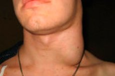
Enlarged cervical lymph nodes, or cervical lymphadenopathy, is not a disease in itself, but a clinical sign. It refers to one or more lymph nodes in the neck becoming larger than normal due to infection, inflammation, an immune response, a tumor, or another cause. In clinical literature, lymph nodes are considered pathologically altered not only by an increase in size but also by changes in density, mobility, and tenderness. In adults, nodes larger than 1 cm are usually considered enlarged, and supraclavicular nodes are considered particularly concerning. [1]
The cervical lymph nodes are part of the immune system. They filter lymph, capture microbes and antigens from the head and neck area, and participate in the immune response. This is why they often react to common upper respiratory infections, tonsillitis, dental problems, viruses, and scalp infections. This reactivity is especially pronounced in children. [2]
In children, enlarged cervical lymph nodes are very common and are often a variant of benign reactive lymphadenopathy following a viral infection. Guidelines from the Royal Children's Hospital Melbourne indicate that cervical lymphadenopathy can be detected in more than one-third of apparently healthy children, and many reactive nodes persist for weeks or even months after a viral infection. [3]
In adults, the situation is different. If an adult develops a persistent neck lump without an obvious infectious cause, especially for more than two weeks, the likelihood of a benign reaction is no longer automatically considered high. The American Academy of Otolaryngology emphasizes that a persistent neck lump in an adult should be considered potentially malignant until proven otherwise. [4]
From a practical standpoint, the key question isn't "Is the lymph node itself dangerous?" but rather "What exactly caused it to enlarge?" Therefore, the correct approach is always based on the cause, duration of existence, patient age, node location, associated symptoms, and examination findings. [5]
Below is a brief table to help understand the general meaning of the findings. The data in it are summarized from clinical guidelines for children and adults. [6]
| Question | Short answer |
|---|---|
| Enlarged cervical lymph node is a diagnosis | No, it's a sign |
| The most common cause in children | Viral infection |
| The most common cause in adults with a short course | Infection or inflammation near a lymph node |
| The most dangerous situation | A persistent painless nodule in an adult with no signs of infection |
| When it is especially alarming | Supraclavicular localization, density, fixation, growth, general symptoms |
Main causes: from a common infection to a tumor
The causes are conveniently divided into four broad groups: infectious, inflammatory and autoimmune, neoplastic, and rarer specific conditions. The most common cause in childhood is viral upper respiratory tract infections. The Royal Children's Hospital Melbourne guidelines explicitly state that viral infection is the most common cause of cervical lymphadenopathy in children, and enlarged reactive nodes can persist for weeks or months. [7]
Infectious causes, in addition to common viruses, include infectious mononucleosis, cytomegalovirus infection, rubella, bacterial tonsillitis, dental infection, inflammation of the salivary glands, cat scratch disease, toxoplasmosis, tuberculosis, and infections caused by nontuberculous mycobacteria. In children with chronic unilateral enlargement of the node with a bluish-violet tint of the skin, nontuberculous mycobacterial infection is especially suspected. [8]
Autoimmune and systemic inflammatory causes are less common, but they should be considered, especially if the lymph nodes are enlarged elsewhere than in the neck. These include systemic lupus erythematosus, juvenile idiopathic arthritis, sarcoidosis, and Kawasaki disease in children. Generalized lymphadenopathy, that is, enlarged nodes in two or more non-adjacent areas, often prompts a search for a systemic cause. [9]
Tumor causes always require special attention. In children, they are rare, but possible, primarily leukemia and lymphoma. In adults, a persistent neck nodule may be a metastasis from cancer of the oral cavity, oropharynx, larynx, thyroid gland, salivary glands, or a manifestation of lymphoma. The American Academy of Otolaryngology notes that most persistent neck masses in adults are neoplasms, not infections. [10]
Finally, there are a group of less obvious causes: drug reactions, scalp and neck eczema, rare immunodeficiencies, common variable immunodeficiency, and combined scenarios where, after an infection, an enlarged node does not return to its original size for a long time. This is why the same symptom should not be automatically interpreted the same way in a 3-year-old child as in a 58-year-old smoker. [11]
The following table helps quickly navigate the causes. It is based on clinical sources for general practice, pediatrics, and internal medicine. [12]
| Group of reasons | The most typical examples |
|---|---|
| Viral | Acute respiratory viral infections, infectious mononucleosis, cytomegalovirus, rubella |
| Bacterial | Streptococcal infection of the oropharynx, staphylococcal lymphadenitis, dental infections |
| Specific infectious | Tuberculosis, nontuberculous mycobacteria, toxoplasmosis, cat scratch disease |
| Autoimmune and inflammatory | Systemic lupus erythematosus, juvenile idiopathic arthritis, sarcoidosis, Kawasaki disease |
| Tumor | Lymphoma, leukemia, metastases of head and neck tumors |
| Other | Drug reactions, dermatological diseases, immunodeficiencies |
How to distinguish a benign reaction from a dangerous signal
The most useful clinical guideline is a combination of five characteristics: age, duration, tenderness, consistency, and location. In a child, a small, mobile, painful nodule after a cold is most often reactive. In an adult, a firm, painless nodule, especially one that persists for more than two weeks without obvious infection, is significantly more suggestive of a tumor. [13]
Tenderness usually indicates acute inflammation, but this is not an absolute rule. Acute bacterial lymphadenitis is often unilateral, painful, and hot, sometimes with redness and fluctuation if an abscess forms. In contrast, tuberculous, lymphomatous, and metastatic nodes are often painless. [14]
Consistency and mobility are also important. According to the American Academy of Otolaryngology and the American Academy of Family Physicians, a firm, fixed, and slightly mobile nodule larger than 1.5 cm, as well as an ulcerated skin overlying it, are associated with an increased risk of malignancy. Nodes that are stuck together may also indicate either a tumor or a specific infection. [15]
Localization is important. Supraclavicular lymph nodes are considered the most worrisome because they are more often associated with malignant and systemic diseases. Posterior cervical nodes can become enlarged with viruses, infectious mononucleosis, tuberculosis, and lymphomas. Submandibular and anterior cervical nodes often respond to infections of the oropharynx, teeth, and salivary glands. [16]
Finally, there are general warning signs: unexplained weight loss, night sweats, persistent fever, weakness, itching, bleeding, and enlarged liver or spleen. When combined with lymph node enlargement, these symptoms greatly increase the likelihood of a systemic disease, including lymphoma, leukemia, tuberculosis, or a severe viral infection. [17]
Below is a table that helps quickly distinguish safer from more dangerous scenarios. It is based on recommendations for the evaluation of lymphadenopathy in children and adults. [18]
| Sign | More often speaks in favor of a reactive process | More often than not, it is alarming in relation to a serious cause. |
|---|---|---|
| Age | Childhood | Age over 40 years |
| Pain | Eat | No |
| Mobility | Good | Limited |
| Consistency | Soft or elastic | Dense or hard |
| Duration | A few days after infection | More than 2 weeks in an adult without improvement |
| Localization | Anterior cervical groups after throat infection | Supraclavicular region |
| General symptoms | No | Weight loss, night sweats, persistent fever |
How doctors examine you: a step-by-step algorithm
The examination begins with a conversation and physical examination. The doctor asks when the nodule appeared, whether it has changed in size, and whether there has been a recent sore throat, runny nose, tooth decay, gum inflammation, a cat scratch, exposure to tuberculosis, taking new medications, weight loss, night sweats, or prolonged fever. At this stage, it is possible to divide some patients into low- and high-risk groups. [19]
The next step is a physical examination. The nodule's size, number, unilateral or bilateral nature, tenderness, density, mobility, skin condition, and the presence of enlarged nodules in other areas are assessed. The oropharynx, teeth, ears, scalp, thyroid gland, salivary glands, and abdomen are also examined. In high-risk adults, a targeted examination of the mucous membranes of the pharynx, larynx, and base of the tongue is essential. [20]
Laboratory tests are not prescribed equally for everyone. If an infection is suspected, a complete blood count, C-reactive protein, serology for Epstein-Barr virus, cytomegalovirus, toxoplasmosis, testing for human immunodeficiency virus, and, if a corresponding history is present, tuberculosis testing may be required. If a hematological disease is suspected, a complete blood count, white blood cell count, blood smear, and further hematological testing are important. [21]
The choice of imaging depends on the age and clinical objective. In children, ultrasound is usually the first method, especially if an abscess or signs of purulent fusion are needed. The American College of Radiology considers ultrasound a generally appropriate first method in children with a neck mass, and a 2024 review of cervical lymph node imaging lists it as the preferred first method for superficial and lateral lymph nodes. [22]
In adults, if a tumor is suspected, the priority shifts to computed tomography (CT) of the neck with intravenous contrast or magnetic resonance imaging (MRI) with contrast. The American Academy of Otolaryngology recommends these methods in adults with an increased risk of malignancy, and the American College of Radiology considers CT and MRI with contrast to be generally appropriate first-line methods in adults with a nonpulsatile neck mass. [23]
If the diagnosis remains unclear after examination and imaging, the next step is morphological verification. In adults, if a tumor is suspected, fine-needle aspiration biopsy is preferred over open biopsy. The American Academy of Otolaryngology guidelines explicitly emphasize that open biopsy is not recommended as a first step because it may impair subsequent treatment and increase the risk of complications. [24]
Below is a step-by-step examination chart. It is based on adult cervical mass guidelines, pediatric guidelines, and imaging guidelines.[25]
| Stage | What are they doing? |
|---|---|
| 1 | They clarify the duration, dynamics, connection with infection and general symptoms |
| 2 | The lymph node and areas of possible infection or tumor sources are examined. |
| 3 | They decide whether the patient belongs to a low or high risk group |
| 4 | Tests are prescribed based on the clinical situation. |
| 5 | In children, they usually start with an ultrasound examination. |
| 6 | In high-risk adults, contrast-enhanced computed tomography or magnetic resonance imaging is more often chosen. |
| 7 | If there is any uncertainty, a fine-needle aspiration biopsy or other morphological verification is performed. |
Treatment: not the lymph node, but the reasons for its enlargement
The main principle of treatment is very simple: an enlarged cervical lymph node itself is not treated as a separate condition. The underlying cause is treated. This is why the same symptom in two patients may require either observation alone, urgent oncological diagnosis, antibiotics, surgery, or anti-tuberculosis therapy. [26]
If the child is feeling well, has recently had a viral infection, and the lymph nodes are small, mobile, and without signs of suppuration, observation is often sufficient. The Royal Children's Hospital Melbourne pediatric guidelines indicate that in an apparently well child, observation and reassurance without examination are often the correct approach. This is especially true if the nodes have been present for a short time and there are no systemic signs of disease. [27]
Symptomatic care includes rest, fluids, age-appropriate pain and fever treatment, and monitoring progress. With reactive viral lymphadenopathy, the node may shrink slowly, and this does not always indicate a complication. Much more important is whether the overall inflammatory background is subsiding and whether new warning signs appear. [28]
In acute bacterial lymphadenitis, especially if the node is unilateral, painful, enlarged, and the overlying skin is reddened, antibacterial therapy is required. Pediatric guidelines recommend oral cephalexin for 7 days with reassessment after 48 hours for uncomplicated bacterial lymphadenitis, and intravenous cefazolin for more severe cases. These regimens are examples; the specific drug choice and duration depend on age, local bacterial resistance, and the severity of the infection. [29]
If fluctuation or a suspected abscess occurs, antibiotics alone may not be sufficient. In this case, an ultrasound is performed, and if a purulent cavity is confirmed, a puncture or surgical incision and drainage is considered. Microbiological examination of the contents helps identify the causative agent and adjust therapy. [30]
When an enlarged cervical lymph node is associated with a source of infection in the teeth, gums, tonsils, ear, or salivary glands, debridement of the source is essential. Without treatment of the underlying dental or otolaryngological problem, the lymph node may shrink only temporarily and then enlarge again. In this situation, treatment of lymphadenopathy without treatment of the source is almost always incomplete. [31]
In the case of tuberculous lesions or infections caused by non-tuberculous mycobacteria, the approach is fundamentally different from the treatment of ordinary purulent lymphadenitis. These require specialized microbiological testing and treatment according to specialized infectious regimens. In children, a chronic, unilateral, mildly painful nodule with a bluish tint of the skin is particularly suggestive of a non-tuberculous mycobacterial infection, while painless nodules with systemic symptoms suggest tuberculosis. [32]
If the cause is infectious mononucleosis, cytomegalovirus infection, or other viral diseases, specific treatment of the lymph node is often not required. In these situations, management is primarily supportive, and the physician's primary goal is to detect complications and avoid prescribing unnecessary antibiotics. Generalized lymphadenopathy combined with liver and spleen enlargement is particularly helpful in understanding the viral nature of the process. [33]
In autoimmune diseases such as systemic lupus erythematosus, juvenile idiopathic arthritis, or other rheumatological conditions, cervical lymph node enlargement only subsides when the underlying disease is controlled. In such cases, treatment is managed not only by a general practitioner or pediatrician, but also by a rheumatologist, and the strategy may include anti-inflammatory and immunomodulatory medications tailored to the underlying disease. [34]
In adults with a nodule suspected of being a tumor, the strategy begins not with a trial course of antibiotics, but with expedited diagnostic testing. The American Academy of Otolaryngology explicitly recommends against routinely prescribing antibiotics unless there is evidence of a bacterial infection. For such patients, contrast imaging and fine-needle aspiration biopsy are the key first steps, rather than waiting for months. [35]
If lymphoma, metastasis from head and neck cancer, thyroid cancer, or another tumor is confirmed, further treatment depends entirely on the histological diagnosis. This may include surgery, radiation therapy, systemic antitumor therapy, and, depending on the specific tumor, modern immune and targeted approaches. However, another crucial point is that an accurate morphological diagnosis is essential before initiating such treatment, which is why open biopsy in adults should not replace the correct diagnostic pathway. [36]
Below is a practical treatment chart based on the causes. It is based on guidelines for children and adults, as well as modern diagnostic algorithms for neck lesions. [37]
| Cause | The basic approach |
|---|---|
| Reactive viral lymphadenopathy | Observation and symptomatic care |
| Acute bacterial lymphadenitis | Antibiotics, re-evaluation soon |
| Lymph node abscess | Visualization, drainage, antibiotics |
| Odontogenic or otolaryngological focus | Treatment of the primary lesion |
| Tuberculosis or non-tuberculous mycobacteria | Specific antimicrobial tactics after confirmation |
| Autoimmune disease | Treatment of the underlying systemic process |
| Suspected tumor | Rapid visualization and morphological verification |
| Confirmed lymphoma or metastasis | Specialized oncological treatment |
Prevention and prognosis
Prevention depends on the cause. For the most common reactive forms, this includes prompt treatment of oropharyngeal and dental infections, oral hygiene, control of chronic inflammation, and careful attention to lingering infections. In children, it's not so much "preventing every nodule" as reducing the risk of severe purulent complications and not overlooking systemic diseases. [38]
It is especially important for adults to promptly seek evaluation of a persistent neck nodule. The American Academy of Otolaryngology guidelines actually suggest that delays worsen outcomes in head and neck tumors. Diagnostic delays of months have been described as a real clinical problem, and shorter delays are associated with worse functional outcomes. [39]
The prognosis in children is generally good, as the underlying cause is often reactive and benign. Guidelines from the Royal Children's Hospital Melbourne clearly state that most cases are benign and self-limiting. However, the prognosis changes dramatically if tuberculosis, a purulent abscess, a systemic disease, or a tumor are involved. [40]
In adults, the prognosis is determined not by the actual enlargement of the node, but by the cause and timing of diagnosis. According to the updated MSD Manual, among undifferentiated cases of lymphadenopathy in adults seen in primary care, cancer occurs in approximately 1%, and in patients over 40 years of age, approximately 4%. This is not a high percentage in absolute terms, but it is significant enough that persistent cervical lymphadenopathy in adults should not be ignored. [41]
Below is a table with practical prognostic guidelines. It is based on guidelines for adults and children and a current professional reference book on lymphadenopathy. [42]
| Situation | What does this usually mean for the forecast? |
|---|---|
| A child after a viral infection, a small mobile node | More often than not, the prognosis is favorable. |
| Acute bacterial lymphadenitis, treatment started on time | Usually a good prognosis |
| Abscess without delayed drainage | The prognosis is usually good |
| A persistent, dense knot in an adult | An oncological route is needed, the prognosis depends on the cause |
| Generalized lymphadenopathy with systemic symptoms | The prognosis is determined by the underlying systemic disease. |
| Late presentation due to malignant cause | Worsens the outcome |
FAQ
Does an enlarged cervical lymph node always indicate something dangerous?
No. In children, it's often a reaction to a viral infection, and many cases resolve on their own. However, in adults, a persistent node without obvious infection requires a more serious evaluation. [43]
Is it possible to simply take an antibiotic and wait?
Not always. Routine antibiotics are not recommended for adults without signs of bacterial infection, as they can only delay proper diagnosis. In children with typical bacterial lymphadenitis, antibiotics are indeed often necessary, but the treatment depends on the clinical presentation. [44]
What lymph node size is considered suspicious?
In general practice, a lymph node larger than 1 cm is considered abnormal, but not only size but also location, density, mobility, and duration are important for evaluation. In adults, the risk is particularly high with a size greater than 1.5 cm, density, fixation, and persistence for more than 2 weeks. [45]
Which is better: ultrasound or CT scan?
It depends on the age and the problem. In children, ultrasound is often the first choice. In adults, if a malignancy is suspected, CT or MRI with contrast is usually needed. [46]
When is a biopsy needed?
It is necessary when the cause remains unclear, there are high-risk features, or imaging does not explain the nature of the nodule. In adults, if a tumor is suspected, the first morphological method is usually fine-needle aspiration biopsy. [47]
Can a lymph node remain enlarged long after a cold?
Yes. Particularly in children, reactive lymph nodes can persist for weeks and months after a viral infection. Not only the duration is important, but also whether the node shrinks, becomes denser, and whether new warning signs appear. [48]
Key points from experts
Melissa A. Pynnonen, MD, MS, Clinical Professor of Otolaryngology and Head and Neck Surgery, University of Michigan. Melissa Pynnonen's professional biography and her contributions to guidelines for the evaluation of neck masses reflect one key finding in contemporary practice: in an adult patient, a persistent neck mass cannot be attributed solely to inflammation for long unless there is a compelling infectious cause. The size of the mass itself is not the determining factor in prognosis, but the speed of diagnosis. [49]
Joseph M. Aulino, MD, associate professor of radiology and radiological sciences, is a neuroradiologist at Vanderbilt University Medical Center. His work on imaging selection criteria underscores another important point: imaging should be tailored to the patient's age and clinical needs. Ultrasound is particularly useful for superficial cervical nodes in children and some adults, while contrast-enhanced CT or MRI are key for deep nodes and suspected tumors. [50]
David Goldenberg, MD, FACS, is professor and chair of the Department of Otolaryngology-Head and Neck Surgery at Penn State Health. His clinical expertise as a head and neck oncology surgeon is particularly relevant to understanding adult cervical lymphadenopathy: sometimes a painless neck nodule is not a local lymph node problem but the initial manifestation of head and neck cancer or lymphoma. The key practical lesson here is that a suspicious neck nodule in an adult requires rapid pathologic evaluation rather than endless courses of treatment "just in case." [51]