Medical expert of the article

New publications
Subluxation of the lens
Last reviewed: 29.06.2025

All iLive content is medically reviewed or fact checked to ensure as much factual accuracy as possible.
We have strict sourcing guidelines and only link to reputable media sites, academic research institutions and, whenever possible, medically peer reviewed studies. Note that the numbers in parentheses ([1], [2], etc.) are clickable links to these studies.
If you feel that any of our content is inaccurate, out-of-date, or otherwise questionable, please select it and press Ctrl + Enter.
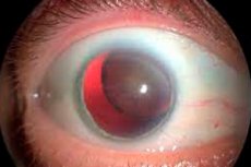
A lens subluxation (or lens dislocation) is a medical condition in which the lens of the eye is partially or completely out of its normal position in the eyeball. This condition can cause severe eye pain and decreased vision. Lens subluxation can be caused by a variety of factors, including trauma, congenital anomalies, abnormalities in the structure of the eye, and others.
If you experience symptoms of lens subluxation, it requires immediate medical intervention. Do not attempt to correct the position of the lens yourself, as this may cause additional damage to the eye. The doctor will examine the eye, possibly with special equipment, and decide how to treat the condition.
Treatment for lens subluxation may include drug therapy, surgery, or other procedures to restore the normal position of the lens and restore vision. It is important to contact an ophthalmologist or emergency medical service immediately if you suspect a lens subluxation to avoid vision loss and complications.
Causes of the lens dislocation
This condition can have a variety of causes, including:
- Eye trauma: Traumatic impact such as a blow, bump, fall, or other injury can cause lens dislocation. This is especially common in athletes and people who work with an increased risk of eye injuries.
- Congenital Anomalies: Some people may have congenital abnormalities of the eye structure that make them more susceptible to lens dislocation.
- Aging: The gradual aging of the body can lead to a deterioration in the elasticity and mobility of the lens, which can increase the risk of lens dislocation.
- Ophthalmologic conditions: Some ophthalmologic conditions, such as Marfan syndrome or Marfan syndrome, may be associated with an increased risk of lens dislocation.
- Eye Surgical Procedures: Someeye surgeries, such as cataract removal or retinal surgery, can increase the risk of lens dislocation.
- Inflammatory diseases of the eye: Certain inflammatory processes within the eye can lead to lens dislocation.
- Heredity: In some cases, heredity may play a role in the development of lens dislocation.
It is important to note that lens dislocation is a serious medical condition that can cause decreased vision and require medical attention.
Pathogenesis
The pathogenesis of lens dislocation includes the following key points:
- Change in the shape of the lens: Normally, the lens has a biological shape that allows it to focus light on the retina. When the lens is dislocated, the lens changes its shape and may move from its normal position inside the eye.
- Distortion of thevisual axis: Moving the lens can disrupt the optical system of the eye, resulting in a distortion of the visual axis. This in turn causes a change in focal length and quality of vision.
- Corneal damage: A dislocated lens can also damage the cornea, the clear outer layer of the eye. This can worsen vision problems and cause additional symptoms.
- Mechanisms of damage: Dislocation of the lens can be caused by various factors such as trauma, natural changes in the structure of the eye (e.g., increase in the size of the ocular globe), disorders associated with aging, and other diseases of the eye.
- Symptoms: A dislocated lens can cause symptoms such as decreased vision, double vision, eye pain, and headaches.
- Treatment: Treatment usually involves surgical correction, during which the lens is returned to its normal position or removed if necessary. Treatment may also include correction of the damaged cornea.
Symptoms of the lens dislocation
Symptoms of lens withdrawal may include:
- Abrupt Visual Impairment: One of the most characteristic symptoms is a sudden and severe visual impairment that can occur suddenly.
- Blurring and double vision: When the lens is dislocated, the image may become blurred or bifurcated.
- Photophobia: Patients often become more sensitive to bright light and may experience pain or discomfort when exposed to bright light.
- Pupildullness or immobility: The pupil may remain dilated and unresponsive to light, which may be a sign of lens dislocation.
- Feelingpressure in the eye: Some patients may feel pressure or discomfort in the eye.
- Headache: A lens dislocation can cause headache, especially if accompanied by other symptoms.
- Floating spotsor turbidity: Floating turbid spots may appear in the visual field.
- Changes in color perception: Patients may notice changes in color perception.
It should be noted that lens dislocation is a medical situation that requires immediate medical attention. If you suspect lens dislocation or have any of the above symptoms, see your doctor or go to the nearest emergency room immediately for evaluation and treatment. This condition can lead to serious complications such as glaucoma and vision impairment, so it is important to get professional medical attention as soon as possible.
A dislocated lens in a child
A lens subluxation (or lens dislocation) is a condition in which the lens, which is normally located inside the eye and serves to focus light on the retina, moves out of its normal position. In children, this condition can occur for a variety of reasons and may require medical intervention.
The main causes of lens subluxation in children may include:
- Trauma: Traumatic exposure, such as a blow, fall, accident, or sports injury, can lead to lens subluxation in children.
- Congenital Anomalies: Some children may have congenital abnormalities of the eye structure that make them more susceptible to lens dislocation.
- Syndromes and Heredity: Certain genetic syndromes may be associated with an increased risk of lens subluxation in children.
- Inflammatory diseases: Certain inflammatory processes within the eye can lead to lens subluxation in children.
If a child has a lens subluxation, it is important to contact an ophthalmologist or emergency medical service immediately for diagnosis and treatment. Diagnosis includes an eye exam and special tests to determine the extent and nature of the subluxation.
Treatment for lens subluxation in children may involve surgery to return the lens to the inside of the eye and prevent additional damage to the eye and loss of vision. The procedure for surgery will depend on your specific situation and symptoms. It is important to discuss all aspects of treatment with your doctor, who will diagnose and prescribe the best treatment for your child.
Stages
This condition can have different stages, depending on how much the lens has moved and how it affects vision. The main stages of lens dislocation may include the following:
- Subluxation (subluxation): In this stage, the lens does not completely move out of its normal location, but may partially move out of the parent capsule that surrounds it. This can cause distortion of vision and patient anxiety.
- Dislocation (complete dislocation): In this stage, the lens is completely out of its normal location and can move inside the anterior chamber of the eye. This causes significant distortion of vision and sometimes blocks the drainage pathway inside the eye, which can lead to increased intraocular pressure.
- Prolapse (passage through the pupil): In this stage, the lens extends beyond the pupil and may be visible to the outside of the eye through the iris. This results in impaired vision and requires immediate medical attention.
The degree and severity of lens dislocation can vary from case to case. Treatment depends on the stage and symptoms. In cases of lens dislocation or lens prolapse, immediate surgical intervention is required to return the lens to the inside of the eye and prevent additional damage to the eye and loss of vision.
Complications and consequences
This refers to ophthalmologic manifestations and can have various complications and consequences depending on the severity and timing of the problem. Here are some of the possible complications:
- Corneal diseases: A dislocated lens can damage the cornea (the clear front part of the eye). This can cause various corneal diseases such as corneal erosion, inflammation of the cornea or scarring.
- Vision impairment: A dislocated lens can cause vision impairment, especially if it is not corrected in a timely manner. This can lead to an impaired focusing of light on the retina and blurred images.
- Glaucoma: In some cases, lens dislocation can increase intraocular pressure and increase the risk of developing glaucoma. Glaucoma is a serious condition that can lead to impaired vision and even blindness if left untreated.
- Cataracts: Long-term effects of lens dislocation on the lens of the eye can contribute to the development of cataracts, resulting in a darkened lens and impaired vision.
- Inflammation and infections: Damage to the lens can increase the risk of inflammation and infection within the eye.
- Astigmatism: A dislocated lens can cause astigmatism, which means that light is not focused on the retina evenly, and this causes distorted visual images.
Diagnostics of the lens dislocation
Diagnosis of lens dislocation is an important step in determining the nature and extent of damage to the eye and developing a treatment plan. Diagnosis usually involves the following procedures and techniques:
- Medical History: The physician collects the patient's medical history, including information about symptoms, previous injuries or surgery, eye diseases, and other medical conditions.
- General ophthalmologic examination: The doctor performs a general ophthalmologic examination of the eye, including vision testing, examination of the structure of the ocular globe, and examination of the anterior and posterior segments of the eye.
- Checking the pressure inside the eye (tonometry): Intraocular pressure measurement may be performed to detect increased intraocular pressure, which may be due to lens dislocation.
- Ultrasound eye examination (ultrasound biomicroscopy): This examination provides a more detailed look at the structures of the eye, including the position and condition of the lens and cornea.
- Computed tomography (CT) or magnetic resonance imaging (MRI): Occasionally, a CT or MRI may be required to visualize ocular structures in more detail and assess the extent of damage.
- Other specialized tests: In some cases, specialized tests and examinations may be needed to further evaluate the condition of the eye and determine the best treatment option. [1]
Differential diagnosis
The differential diagnosis of lens output involves identifying the condition and distinguishing it from other diseases or conditions that may mimic the symptoms. Some of the possible differential diagnoses include:
- Glaucoma: Glaucoma is a condition characterized by an increase in intraocular pressure, which can cause blurred vision, eye pain and headaches. Glaucoma should be ruled out because high intraocular pressure can be dangerous.
- Cataract: A cataract is a darkening of the lens that can cause vision changes. It can also mimic the symptoms of lens withdrawal. Determining if a cataract is present may require an eye exam by a doctor.
- Migrainewith aura: Migraine with aura can cause temporary changes in vision, including splitting, flickering, and blurring. It is important to distinguish this condition from lens withdrawal.
- Macular Degeneration: Macular degeneration is a condition in which the macula (part of the retina) degenerates, which can cause a deterioration of central vision.
- Acquired retinal diseases: Various retinal diseases can cause changes in vision and can mimic the symptoms of lens withdrawal.
- Traumatic injuries to the eye: Trauma to the eye can cause changes in the position of the lens or other abnormalities, which can also mimic symptoms.
A comprehensive eye examination by an ophthalmologist is necessary to make a differential diagnosis and determine the exact cause of symptoms. This may include examination of the fundus of the eye, measurement of intraocular pressure, and additional tests such as eye ultrasound, optical coherence tomography (OCT) or others.
Treatment of the lens dislocation
Treatment for lens dislocation, also known as lens nucleus dislocation or lens dislocation, can vary depending on the extent of the dislocation and the presence of complications. It is important to see an ophthalmologist as soon as possible to diagnose and treat this condition. The following are common treatment options:
- Restoring the lens to its normal position (lens refraction): This process may be performed by an ophthalmologist using specialized instruments. The lens is returned to its place in the eye. It is important to have this procedure performed by an experienced professional.
- Fixation of the lens: After refraction of the lens, fixation of the lens may be necessary to prevent it from dislocating again. This can be accomplished in a variety of ways, including the use of sutures or other methods.
- Monitoring and treating complications: Your doctor will monitor the condition of your eye after lens repair and treat any complications such as inflammation, infection, or increased intraocular pressure.
- Glassesor contact lenses: In some cases, especially if the lens has been removed or cannot be restored, the patient may need glasses or contact lenses to correct vision.
- Surgery: In cases where the lens cannot be repaired or there are serious complications, surgical intervention such as implantation of an artificial lens (phacoemulsification and intraocular lens) or other surgical procedures may be required to restore vision. [2]
Surgery for lens dislocation
Surgery to restore the correct position of the lens (treatment of a lens subluxation) may be necessary, especially if the lens is completely out of its normal location and this causes vision impairment. This surgery is usually performed by an ophthalmologist and may be called lens repair surgery or lens surgery.
The surgical procedure may vary depending on the specific situation and the degree of lens dislocation. The basic steps of surgery may include the following:
- Patient Preparation: Instructions may be provided to the patient on how to prepare for surgery, including temporary medication and eye preparation.
- Anesthesia: The eye is usually anesthetized to ensure the patient's comfort during surgery.
- Access to the lens: Through a small incision near the cornea or sclera, which may be called a sclerocorneal incision, the surgeon accesses the lens.
- Lensrepair: The surgeon manipulates the lens and returns it to its normal position inside the eye. In some cases, if the lens is damaged or cannot be repaired, it may be removed (lens extraction).
- Completion of the surgery: After the lens is repaired or removed, the surgeon closes the incision and may use sutures or glue to secure the tissue. The surgery can be performed using either microsurgical instruments or lasers.
- Aftercare: The patient may require the use of medicated drops to prevent infection and aid in healing.
After surgery, the patient should follow the doctor's recommendations for care and medications to ensure optimal recovery. The success of the surgery depends on many factors, including the extent of damage to the lens and the general condition of the eye.
Treatment for lens dislocation should be individualized and depend on each patient's specific situation.