
New publications
Subcutaneous adipose tissue: structure, functions, differences from visceral fat and clinical significance
Last updated: 27.03.2026

All iLive content is medically reviewed or fact checked to ensure as much factual accuracy as possible.
We have strict sourcing guidelines and only link to reputable media sites, academic research institutions and, whenever possible, medically peer reviewed studies. Note that the numbers in parentheses ([1], [2], etc.) are clickable links to these studies.
If you feel that any of our content is inaccurate, out-of-date, or otherwise questionable, please select it and press Ctrl + Enter.
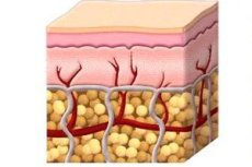
Subcutaneous fat is the deepest layer of the skin, lying beneath the dermis and connecting the skin to the underlying muscles, fascia, and other tissues. In anatomical sources, this layer is also described as the hypodermis or subcutaneous tissue. It consists primarily of fat and loose connective tissue and also contains blood vessels, lymphatic structures, nerves, and some immune system cells. [1]
Viewing this layer solely as a "fat reserve" is overly simplistic. Modern reviews show that subcutaneous fat is not a passive cushion, but an active tissue with mechanical, metabolic, endocrine, and immune functions. It protects against impact, helps retain heat, serves as an energy storage site, and simultaneously plays a role in regulating metabolism and immune responses. [2]
The structure of subcutaneous fat tissue also varies across the body. Its thickness varies depending on the body region, gender, age, genetics, and hormonal levels. Clinical studies indicate that this layer is particularly thin in the eyelids and external genitalia and much thicker in the abdomen and buttocks. This is not simply an anatomical detail: the distribution of subcutaneous fat affects body contours, heat exchange characteristics, and some metabolic risks. [3]
It's also important to understand that subcutaneous adipose tissue contains more than just mature fat cells. It also contains stromal cells, fibroblasts, vascular elements, macrophages, lymphocytes, and other cell populations that influence inflammation, healing, and local metabolism. A 2024 review emphasized that subcutaneous adipose tissue contains a significant reservoir of immune and stromal cells that influence not only the skin but also systemic processes. [4]
In everyday medicine, this tissue is very important. It is through it that subcutaneous injections are administered, and it is precisely this tissue that doctors evaluate for lipodystrophy, lipedema, panniculitis, lipomas, obesity, and age-related tissue loss. Therefore, subcutaneous fat is not a "secondary layer beneath the skin," but a fully-fledged anatomical and functional system. [5]
The summary table below is compiled from data from anatomical reviews, clinical materials and modern reviews on adipose tissue. [6]
| Component | What's included | Practical significance |
|---|---|---|
| Fat lobules | Adipocytes separated by connective tissue septa | Energy storage, cushioning |
| Connective tissue | Collagen and elastic fibers, septa | Support, fixation, mechanical strength |
| Blood vessels | Arteries, capillaries, veins | Tissue nutrition, metabolism |
| Lymphatic vessels | Lymphatic capillaries and vessels | Fluid drainage and participation in immune defense |
| Nervous elements | Sensory nerve fibers | Feeling of pressure, pain, temperature |
| Immune cells | Macrophages, lymphocytes, mast cells | Inflammation, protection, healing |
What functions does subcutaneous fat perform?
Its most well-known function is energy storage. White adipose tissue, which comprises the bulk of subcutaneous fat, serves as the largest energy depot in the human body. It accumulates lipids and, when needed, participates in their mobilization, helping to maintain energy balance between meals and during periods of calorie deficit. [7]
Equally important is its mechanical role. Subcutaneous fat acts as a shock absorber: it softens impact, redistributes pressure, and allows the skin to glide freely over underlying tissues. Anatomical sources emphasize that it connects the skin to muscles and bones, and also protects organs, muscles, and bones from traumatic impacts. [8]
The third major function is thermal insulation. Adipose tissue helps reduce heat loss and is involved in maintaining temperature homeostasis. Clinical materials and anatomical reviews describe the hypodermis as a layer that protects against cold and helps conserve heat, and as the thickness of this layer decreases, for example with age, its protective ability also diminishes. [9]
The fourth function is endocrine. Adipose tissue has long ceased to be viewed as a silent reservoir of calories. Modern sources describe it as an endocrine organ that secretes leptin, adiponectin, resistin, estrogens, and a whole spectrum of cytokines and other signaling molecules. These substances influence appetite, insulin sensitivity, inflammation, lipid metabolism, and overall metabolic stability. [10]
The fifth function is immune and regenerative. Subcutaneous adipose tissue is involved in immunoregulation, skin protection from infection, and wound healing. A 2024 review emphasizes that processes in this tissue are involved in skin healing, inflammatory skin diseases, and systemic immunometabolic responses. The role of adipocytes and cells in this area in regulating inflammation at the wound edge and tissue remodeling is also described. [11]
Finally, this tissue also plays a practical medical role as a drug administration site. The subcutaneous route is widely used for insulin, heparin, some biological drugs, and infusion therapy. It is valued for its relative simplicity, good tolerability, and the ability to gradually absorb the drug from the subcutaneous space into the bloodstream and lymphatic system. [12]
The table below summarizes the main functions of subcutaneous fat and their clinical significance.[13]
| Function | How is it implemented? | Why is this important in the clinic? |
|---|---|---|
| Energy | Accumulation and mobilization of lipids | Associated with nutrition, body weight, insulin resistance |
| Depreciation | Shock and pressure relief | Protects tissues from mechanical injury |
| Thermal insulation | Reducing heat loss | Important for cooling and age-related tissue thinning |
| Endocrine | Secretion of adipokines and cytokines | Affects metabolism and inflammation |
| Immune | Involvement of immune cells and mediators | Important for infection, inflammation, and healing |
| Drug depot layer | Absorption of subcutaneously administered drugs | The basis for many injectable treatment regimens |
How is subcutaneous fat different from visceral fat?
The key anatomical difference is simple: subcutaneous fat is located beneath the skin, while visceral fat surrounds the internal organs. Visceral fat is located primarily in the omentum and mesentery and drains into the hepatic portal vein system, so its metabolic impact on the liver and overall metabolism is particularly significant. This is why two people with the same body weight can have very different cardiometabolic risks. [14]
According to current data, visceral fat is, on average, more closely associated with an unfavorable metabolic profile than subcutaneous fat. Endocrinology reviews indicate that increased visceral and upper body fat are associated with higher triglycerides, lower high-density lipoprotein cholesterol, and greater insulin resistance. Subcutaneous fat in the legs, on the other hand, has been associated with a more favorable lipid profile in some studies. [15]
However, one shouldn't draw the sweeping conclusion that all subcutaneous fat is always "good." Research shows that within the subcutaneous fat depot, there are different zones with different behaviors. In the abdominal region, superficial and deep layers are distinguished, separated by a fascial septum. Deeper abdominal subcutaneous fat has been shown in a number of studies to have more unfavorable associations with hepatic steatosis and metabolic disorders than superficial fat. [16]
At the same time, there is also a protective potential for subcutaneous fat storage. Research from the Framingham Project and data on lipodystrophy support the idea that the ability to safely store excess energy in subcutaneous adipose tissue may partially protect against ectopic fat deposition in the liver, muscles, and other organs. When such a reserve is depleted or lost, metabolic complications often worsen. [17]
This is why modern medicine prioritizes not only the total volume of fat but also its distribution. Subcutaneous fat is neither uniquely harmful nor uniquely beneficial. Its value depends on the area, depth, tissue capacity for normal expansion, the level of inflammation, and whether excess lipids are shifting to visceral and ectopic depots. [18]
The table below summarizes the main differences between subcutaneous and visceral adipose tissue.[19]
| Sign | Subcutaneous adipose tissue | Visceral adipose tissue |
|---|---|---|
| Where is it located? | Under the skin | Around the internal organs |
| The main role | Energy reserve, mechanical protection, thermal insulation | Metabolically active intra-abdominal depot |
| Connection with the liver | More indirect | More direct through the gate system |
| Metabolic risk | On average, it is lower, but it depends on the zone and structure. | On average, higher |
| Internal heterogeneity | Yes, especially in the abdominal area | There is also one, but clinically it is more often assessed as a single visceral depot |
| Potential protective effect | Possible with preserved ability to safely store lipids | Expressed much less strongly |
How subcutaneous fat changes with age, gender, and body weight
The distribution of subcutaneous fat tissue varies significantly depending on gender and hormonal levels. Clinical data and metabolic studies indicate that with higher estrogen levels, fat tissue is more likely to accumulate in the buttocks, thighs, and pelvis, while with higher androgen levels, it is more likely to accumulate in the abdomen, shoulder girdle, and upper torso. This is one reason why the same body mass index in different people does not necessarily mean the same risk profile. [20]
With age, subcutaneous adipose tissue doesn't simply "get smaller." It changes its cellular composition, adipocyte size, and immune status. A 2024 study on human abdominal white adipose tissue found that aging is associated with an increase in adipocytes, an increase in the proportion of lipid-associated macrophages and mast cells, and increased immune and fibrotic signaling. This explains why age-related changes in adipose tissue affect not only appearance but also metabolism. [21]
At the same time, the hypodermis itself, as a structural layer, thins. Clinical data from the Cleveland Clinic indicate that with age, the hypodermis shrinks, and the skin begins to sag more because there is less volume and connective support between it and the underlying tissues. Externally, this manifests as a decrease in turgor and age-related loss of soft tissue volume. [22]
In obesity, changes are also not limited to simple calorie accumulation. If subcutaneous fat tissue is capable of expanding relatively physiologically, it partially absorbs the excess energy. If its adaptive capacity is limited, inflammation, fibrosis, and lipid redistribution to the liver and muscles begin to increase. This scenario is clearly evident in lipodystrophy, where a deficiency in subcutaneous fat stores leads to severe metabolic disturbances. [23]
Patients sometimes mistakenly believe that localized subcutaneous fat removal automatically improves metabolism. However, surgical sources emphasize that liposuction is primarily a contouring procedure, not a weight-loss procedure. Earlier metabolic observations also showed that subcutaneous fat removal does not necessarily produce the same effect on glucose and lipids as visceral fat reduction. This further confirms that it's not just fat volume that matters, but its biology and distribution. [24]
The summary table below shows how subcutaneous fat changes in different physiological states. [25]
| State | Typical changes |
|---|---|
| Young age | More preserved structure and volume, better tissue restoration |
| Aging | Thinning of the hypodermis, enlargement of adipocytes, and increased inflammatory and senescent signals |
| Female hormonal profile | More accumulation in the hips and buttocks area |
| Male hormonal profile | More accumulation in the abdominal and upper torso area |
| Obesity | Expansion of the depot, possible inflammation and fibrosis |
| Lipodystrophy | Loss of subcutaneous fat with risk of ectopic lipid accumulation |
When subcutaneous fat becomes a source of clinical problems
One of the most discussed pathologies is lipedema. This condition is characterized by disproportionate and symmetrical accumulation of adipose tissue, most often in the lower extremities, often accompanied by pain, easy bruising, and a poor response to normal weight loss. StatPearls and Endotext emphasize that lipedema is a disease of the subcutaneous fat tissue and is more common in women. [26]
Another major class of conditions is lipodystrophies, in which subcutaneous fat tissue is partially or almost completely reduced. These diseases are important not only visually but also metabolically. Endotext notes that when subcutaneous fat stores are depleted, excess energy begins to be stored in the liver and other organs, contributing to severe hypertriglyceridemia, insulin resistance, and hepatic steatosis. [27]
The third major group is panniculitis, which refers to inflammation of the subcutaneous fat. StatPearls describes panniculitis as a heterogeneous group of conditions involving inflammation of the subcutaneous fat tissue, which clinically presents as painful subcutaneous nodules or plaques, most commonly on the lower extremities. A deep biopsy is often necessary for an accurate diagnosis, as different types of panniculitis can appear very similar. [28]
A very common benign finding is a lipoma. It is a soft, usually painless subcutaneous tumor of mature fat cells. Most lipomas are superficial, grow slowly, and do not require urgent intervention unless they cause pain, rapid growth, functional problems, or cosmetic discomfort. However, if they are growing rapidly, are dense, attached to deep tissue, or have an atypical location, the doctor should rule out more serious lesions. [29]
There are also rare but important diseases of painful adipose tissue, such as Dercum's disease. StatPearls describes it as a rare chronic condition characterized by multiple painful subcutaneous lipomas, most often found in middle-aged women. Clinically, this condition is often confused with lipedema, fibromyalgia, or regular obesity, which delays proper patient management. [30]
Painful subcutaneous nodules, rapidly growing growths, sudden asymmetry, a pronounced tendency to bruise, persistent swelling, areas of redness and heat, unexplained loss of subcutaneous fat, and a combination of subcutaneous fat changes and metabolic disorders are considered reasons to consult a doctor. In these situations, it's no longer a matter of body type, but a possible disease of the subcutaneous fat tissue. [31]
The summary table below helps to differentiate the major clinical problems of subcutaneous fat.[32]
| State | What is typical |
|---|---|
| Lipedema | Symmetrical, painful accumulation of fat, most often on the legs, more common in women |
| Lipodystrophy | Partial or severe loss of subcutaneous fat with metabolic complications |
| Panniculitis | Painful inflammatory nodules in the subcutaneous fat |
| Lipoma | A soft, usually painless, benign growth |
| Dercum's disease | Multiple painful subcutaneous lipomas and chronic pain |
| Age-related atrophy of the hypodermis | Thinning of the subcutaneous layer and loss of soft tissue volume |
FAQ
Are subcutaneous fat and fat under the skin the same thing?
In a practical sense, yes, but it's more accurate to say that subcutaneous fat is not just the fat cells themselves, but the entire layer, including fat lobules, connective tissue, blood vessels, nerves, lymphatic structures, and immune cells. [33]
Is subcutaneous fat always less dangerous than visceral fat?
On average, visceral fat carries a higher metabolic risk, but subcutaneous fat tissue is also heterogeneous. Deep abdominal subcutaneous fat may behave less favorably than superficial fat, and the overall significance depends on the distribution and function of the tissue. [34]
Why is fat in the hips and buttocks often considered metabolically "calmer"?
Because a number of studies link increased subcutaneous fat in the legs and gluteofemoral region to a more favorable lipid and carbohydrate profile compared to a predominance of visceral and upper body fat. [35]
Can subcutaneous fat become inflamed?
Yes. Panniculitis is a group of conditions in which subcutaneous fat becomes inflamed. Clinically, this often manifests as painful nodules, redness, and sometimes systemic symptoms. [36]
Why are metabolic disturbances so severe in lipodystrophy, despite low body fat?
Because the problem isn't simply a "low amount of fat," but a lack of adequate subcutaneous fat stores for safe lipid storage. As a result, fat begins to accumulate in the liver and other organs, and adipokine levels, especially leptin, decrease. [37]
Does liposuction treat obesity as a disease?
No. Surgical sources emphasize that liposuction is primarily a body contouring method, not a treatment for obesity as a systemic metabolic disease. [38]
Why does skin become more saggy with age, even without significant weight loss?
One reason is a decrease in the volume of the hypodermis and a weakening of the supporting role of subcutaneous fat. This is compounded by changes in the dermis, collagen, and skin elasticity. [39]
When do changes in subcutaneous fat require investigation?
When pain, nodules, persistent swelling, rapidly growing masses, unusual asymmetry, multiple bruises, significant loss of subcutaneous fat, or a combination of these signs with severe metabolic disorders appear. [40]
Key points from experts
Philip Scherer, PhD, is a professor of internal medicine at the University of Texas Southwestern Medical Center. His research on fat cells and the discovery of adiponectin have radically changed the understanding of adipose tissue: from a passive reservoir of calories, it has become a recognized endocrine organ that influences energy balance and the immune system. This is a key point for subcutaneous fat: the importance of this tissue is determined not only by its volume but also by the quality of its signals. [41]
Matthias Blüher, MD, is Professor of Clinical Obesity Research at the University of Leipzig, Senior Physician in the Department of Endocrinology and Nephrology at the University Hospital Leipzig, and Director of the Institute for Metabolism, Obesity, and Vascular Research. His research focuses on adipose tissue dysfunction and fat distribution as mechanisms of type 2 diabetes and cardiovascular disease. The practical implication of his work is that fat assessment should consider not only body mass but also the depot, depth, and biological quality of adipose tissue. [42]
S. Ronald Kahn, MD, is the Academic Director and Chief of the Section of Integrative Physiology and Metabolism at the Joslin Diabetes Center and Professor of Medicine at Harvard Medical School. His research interests include the development and function of adipose tissue and its impact on insulin sensitivity and the risk of metabolic disease. His scientific position supports the current view of subcutaneous adipose tissue: it is not a secondary anatomical layer, but rather part of an interorgan communication system important for metabolism. [43]