Medical expert of the article

New publications
Muscle stretching in lumbosacral spine osteochondrosis
Last reviewed: 06.07.2025

All iLive content is medically reviewed or fact checked to ensure as much factual accuracy as possible.
We have strict sourcing guidelines and only link to reputable media sites, academic research institutions and, whenever possible, medically peer reviewed studies. Note that the numbers in parentheses ([1], [2], etc.) are clickable links to these studies.
If you feel that any of our content is inaccurate, out-of-date, or otherwise questionable, please select it and press Ctrl + Enter.
It is recommended to introduce this methodical technique into the massage procedure immediately after preparing the corresponding muscle with massage techniques (stroking, rubbing, kneading and vibration).
Stretching techniques are used when the elastic properties of the tissues of the locomotor apparatus and skin deteriorate, and muscle tone increases excessively. Their intensity is dosed by the degree of active tension of the muscles that produce stretching, special starting positions. The stretching effect can be increased by additional efforts of the doctor (massage therapist). With the systematic use of stretching, morphological restructuring and improvement of the elastic properties of pathologically altered tissues that cause deformation occur.
ATTENTION! When stretching atrophic (weakened), degeneratively altered and denervated muscles, there can easily be a risk of overstretching them, subsequent deterioration of function (in particular, decreased strength) and slowing down of the processes of normalization of activity.
 [
[ Muscle stretching technique
Abdominal muscles ("pseudovisceral pain")
Trigger points of the abdominal muscles cause suffering not only from reflected pain, but also from induced visceral disorders. Symptoms of damage to internal organs caused by myofascial TPs often complicate diagnosis. Unilateral TPs often cause pain on both sides. In this case, patients usually complain of "burning" in the abdomen, "overflow", "bloating", "swelling", "gases", etc.
- Oblique abdominal muscles. Active TPs of the upper portion of the external oblique abdominal muscle, located anterior to the ribs, cause heartburn and other symptoms usually characteristic of a hernia of the esophageal opening of the diaphragm. TPs localized in one of the three muscle layers of the lower lateral abdominal wall reflect pain to the groin area. Active TPs causing pain along the upper edge of the pubic bone and in the lateral half of the inguinal ligament can be the cause of increased excitability of the detrusor and spasm of the sphincter of the bladder, which is manifested by frequent urination or urinary retention.
- Rectus abdominis muscle. In the upper section, TT located at this level, both on the right and on the left, reflect girdle pain. When TT is localized in the periumbilical region, cramping intestinal colic is not uncommon (Kellgrent J., 1977; Murray J., 1975). Lateral TT can provoke diffuse abdominal pain, which intensifies with TT movements; muscles located in the lowest sections reflect pain bilaterally to the sacroiliac and lumbar regions (Fig. 6.31, b).
Rectus Abdominis Stretching Technique
The patient's initial position is lying on his back (a cotton-gauze roll is placed under the lower back), hands are placed under the head, legs are lowered down, feet are on a stool. The difference between the levels of the table and the stool should be approximately 60 cm. The patient, arching his back, takes a deep breath. At this time, the muscle is stretched.
External Oblique Abdominal Muscle Stretching Technique
The patient's initial position is lying on the healthy side, the shoulder is abducted backwards, to the plane of the couch. In this case, the thoracolumbar spine is rotated, as in stretching the anterior serratus muscle.
To inactivate myofascial TP, it is advisable to include the following exercises in exercise therapy sessions:
- abdominal breathing, as it is useful for stretching the oblique abdominal muscles;
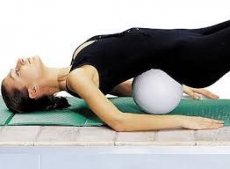
- pelvic lift. The exercise is aimed at stretching the lumbar muscles and training the abdominal muscles.
The patient's initial position is lying on his back, one hand is placed in the pubic symphysis area, the other in the epigastric region, the legs are bent at the knees and hip joints. The patient "presses" the lumbar region on the surface of the couch, while the abdominal muscles contract, aligning the spine (the hands come together). The subsequent movement: lifting the pelvis with a straight back (the hands touch each other). Then the patient returns to the initial position; the exercise is supplemented with breathing and relaxation exercises.
- The sit-lie exercise involves tensing the elongated rectus abdominis muscle, not the shortened one. Stages of the exercise:
- First, the patient slowly lies down on his back from a sitting position (legs bent at the knees and hip joints). Pauses between movement phases should be within 15-30 seconds (isometric muscle tension);
- bending the trunk from the patient's initial position - lying on his back. The patient lifts his head from the plane of the couch, then the shoulder girdle, shoulder blades, without lifting his lower back;
- transition from the patient's initial position - lying down to initial position - sitting. To increase tension, hands should first be placed on the hips, then on the stomach, on the chest and, finally, behind the head.
Stretching technique.
- The patient's initial position is sitting on a chair. The doctor uses his hand to help the patient bend his torso forward while simultaneously rotating it; the patient turns his face in the same direction.
- Corrective exercises to help stretch muscles:
- stretching of the paravertebral muscles of the lower back;
- stretching of the thoracic and lumbar paravertebral muscles in an aquatic environment.
Back muscles
- Superficial paravertebral muscles. The most frequently active TPs appear in the longissimus and iliac-costal muscles of the chest. The latter reflects pain mainly upwards, and the iliac-costal muscles of the lumbar region and the longissimus of the chest - mainly downwards.
Symptoms of damage to the left iliocostalis muscle of the chest imitate the signs of angina pectoris, and the right or both - the picture of pleurisy (Yann C. et al., 1978). Lange M. (1931) described damage to the muscle that straightens the spine at the level of the lower back as a frequent cause of "lumbago" and sacral pain. Later, many patients with referred pain emanating from myalgic areas or painful points in the muscle that straightens the spine were reported in muscular rheumatism.
Muscle stretching technique.
- Patient's initial position: sitting on a chair, feet shoulder-width apart, arms down, torso leaning forward.
- Patient's initial position - sitting on the couch, legs straight. The patient should touch his toes with straight arms.
When performing a stretching procedure, the doctor uses his hand to help the patient perform the exercise, thereby increasing the bending movement.
- Deep paravertebral muscles. Deep muscles more often than superficial ones reflect pain to the anterior abdominal wall. Involvement of the deepest paravertebral rotator muscles in the process causes pain along the midline of the back and reflected pain during percussion along the adjacent spinous processes. And only deep palpation allows us to determine from which side the pain is coming.
ATTENTION! It is advisable to perform the movement on a long exhalation.
Thigh muscles
1. Hip flexor muscles
- Muscle tensor fasciae femoris - active TT are located in its upper third. The pattern of referred pain is detected along the lateral surface of the thigh.
- Pectineus muscle - active TT is projected in the inguinal region. Pattern of referred pain - medial surface of the upper third of the thigh.
- Quadriceps femoris (rectus) - active TPs are diagnosed at the muscle attachment sites. The pattern of referred pain is projected along the muscle and is concentrated in the knee joint area.
- Iliolumbar muscle - active TT are located in the groin area, navel area and upper third of the quadriceps muscle.
Technique for stretching the hip flexor muscles.
- Patient's initial position - lying on the stomach. Alternate raising of straight legs. The affected limb is raised with the help of the doctor's hands.
- Patient's initial position: kneeling at the gymnastic wall, holding the bar with his hands. Maximum extension of the affected leg at the hip joint, without lifting the toe off the floor.
- Patient's initial position - standing on all fours, the affected leg is maximally extended with the support on the toe (the leg and the body form one straight line). Bending the healthy leg to the limit in the hip and knee joints while simultaneously sliding the affected leg back.
2. Hip extensors
- Gluteus maximus muscle.
- Gluteus medius muscle.
A) Technique for stretching the gluteal muscles.
The biceps femoris, semimembranosus and semitendinosus muscles of the thigh are active TT located in the middle third of the back of the thigh. The pattern of referred pain is projected in the upper third of the thigh.
- Patient's initial position - lying on the back, legs straight, arms along the body. Slowly bend the leg at the hip and knee joints, then bend the other leg and use the hands to pull them to the chest (the hands are positioned in a "lock");
- The patient's initial position is the same, but with one hand the doctor bends the patient's head and shoulders forward, while simultaneously applying light pressure on the legs with the other hand.
B) For passive stretching of the muscle fibers of the gluteus medius muscle in the patient's starting position - lying on the healthy side, it is necessary to bring the thigh bent at the hip joint.
- The patient's initial position is lying on his stomach, the leg is bent at the hip and knee joints. The doctor fixes the patient's pelvis with one hand and rotates the leg outward with the other.
Technique for stretching the posterior thigh muscles.
- Patient's initial position - lying on the back. Bend the leg at the hip and knee joints, then use your hands to slowly straighten it, increasing the angle of elevation.
3. Adductor muscles of the thigh. Active TT are localized in the middle third of the inner surface of the thigh.
Technique for stretching the adductor muscles of the thigh.
- Patient's initial position - lying on the back. Legs spread apart;
- patient's initial position - standing sideways to the gymnastic wall on the healthy leg, the affected leg is moved to the side, the foot is on the 3rd - 4th rail - squat, bending the healthy leg;
- patient's initial position - sitting on the bed, holding onto the backrest crossbar with his hands - simultaneously spreading the legs to the sides, gradually lowering them from the bed, the patient seems to sit astride the bed;
- The patient's initial position is lying on his back, legs straight. The doctor fixes the healthy leg in the lower third of the thigh with one hand, and moves the affected leg to the side with the other hand.
Calf muscles
Gastrocnemius muscle. Active TPs are located in the upper third of the shin. The pattern of referred pain covers the entire muscle mass and part of the plantar surface of the foot.
Technique for stretching the calf muscles.
- The patient's initial position is lying on his back, legs straight. The doctor, grasping the lower third of his shin with his hand, performs dorsal flexion of the foot with the other hand, first with the leg bent at the knee and hip joints, then with the leg straight.