Medical expert of the article
New publications
Sequestrectomy
Last reviewed: 29.06.2025

All iLive content is medically reviewed or fact checked to ensure as much factual accuracy as possible.
We have strict sourcing guidelines and only link to reputable media sites, academic research institutions and, whenever possible, medically peer reviewed studies. Note that the numbers in parentheses ([1], [2], etc.) are clickable links to these studies.
If you feel that any of our content is inaccurate, out-of-date, or otherwise questionable, please select it and press Ctrl + Enter.

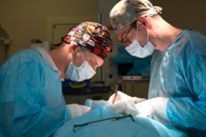
Sequestrectomy is a type of necrectomy, the essence of which is the removal of a sequestrum - a piece of dead tissue (e.g. Necrotized bone segment in osteomyelitis). Sequestrectomy is performed after the sequestrum has completely separated from normal tissue and a sequestral capsule has formed. [1]
Most often, sequestrectomy is not a stand-alone intervention, but a component of a more extensive operation to eliminate the primary pathologic process (for example, in chronic osteomyelitis).
Indications for the procedure
In most cases, sequestrectomy is performed for chronic purulent-necrotic bone lesions, for example, in chronic osteomyelitis, when the formation of fistulous passages, sequestrations, false joints, and cavities is noted. Surgery is indicated if there are frequent recurrences, malignancy of the affected area occurs, or other pathological processes develop due to the presence of a chronic infectious focus. [2]
Sequestrectomy may be indicated at any stage of osteomyelitis (both acute and chronic) if irreversible bone destruction occurs.
Other possible indications for sequestrectomy surgery include:
- Ulcerative processes that develop against the background of a neglected stage of osteomyelitis;
- Formation of fistulas, pustules, as a consequence of internal infectious processes with an acute course;
- Malignant tumors that spread to bone tissue and lead to bone destruction;
- Dysfunction of internal organs, which is due to prolonged intoxication due to osteomyelitis.
Preparation
Sequestrectomy, like any other intervention, requires special preparatory measures. Preliminary diagnostics is carried out, which may include:
- Consultations with a dentist, otolaryngologist, maxillofacial or thoracic surgeon, vertebrologist, orthopedist (depending on the location of the pathological focus);
- X-ray examination of the affected area in 2-3 projections, and if there is a lack of information - the connection of magnetic resonance or computed tomography;
- Fistulography with injection of contrast agent into the fistula.
If general anesthesia is to be used during the sequestrectomy, then additional administration:
- A consult with a therapist, an anesthesiologist;
- Electrocardiography;
- General clinical blood and urine tests;
- Blood chemistry, coagulogram;
- Tests to identify the infectious agent.
Other diagnostic procedures may also be used according to individual indications.
Preoperative preparation for sequestrectomy may include therapeutic measures:
- Inhibition of the inflammatory process in the area of the pathological focus (antiseptic lavage, treatment of fistulous passages and cavities with proteolytic enzymes);
- Skin sanitation in the area of the proposed surgical field;
- Strengthening of immunobiological activity of the organism;
- Normalizing the function of vital systems.
Radical surgery is the main prerequisite for the treatment of sequestrations. It may include both sequestrectomy and fistula excision, bone trepanation with opening of the osteomyelitic sequestral box, cavitary removal of dead granulation and suppurative walls to healthy tissue, repeated cavity sanation with antiseptics. [3]
Technique of the sequestrectomies
Among the possible surgical interventions for chronic osteomyelitis, the most common are:
- Bone resection;
- Osteoperforation;
- Sequestrectomy.
Sequestrectomy for osteomyelitis is subdivided, in turn, into these variants:
- Sequestrectomy with osteoperforation;
- Sequestrectomy with blood clot grafting (proximal or distal);
- Sequestrectomy with bone grafting.
Bone cavity grafting is possible with autogenous, heterogeneous, homogeneous tissue or alloplastic material.
A cavity bone filling is performed:
- With implantable fillings (sponge, porous materials);
- Blood clots with antibiotics (use possible on small cavities);
- Muscle flap, shredded muscle, cartilage, bone, or bone chips.
In patients with posttraumatic chronic osteomyelitis complicated by pseudarthrosis, sequestrectomy is supplemented by false-joint resection with further bone repositioning. [4]
Surgery is usually performed against the background of prolonged therapy, which involves the elimination of purulent inflammation and restoration of impaired motor function. Sequestrectomy is performed in compliance with the following principles:
- To ensure the exit of purulent contents;
- Tissue excision, which allows qualitative removal of the sequestrum without damaging it;
- Excision of the fistula tracts;
- Preservation of newly formed normal bone tissue to ensure bone regeneration processes.
Sequestrectomy is performed using general or local anesthesia. The incision can be made either through the fistula canal or in another convenient place in the area of healthy tissues. To clarify the localization of the sequestrum and purulent-inflammatory foci, information obtained during radiography and fistulography is used.
The surgeon dissects the skin, subcutaneous fatty tissue, fascia, muscles, after which he exposes the area of periosteum and excises the superficial foci together with it. If there are deep-seated foci, the doctor performs dissection and peeling of the periosteum.
After removing all dead tissues, the surgeon sutures the wound, installing a catheter for washing and drainage with antiseptics and antibacterial drugs. The wound is bandaged, if necessary, immobilization with a bandage made of plaster or plastic. After a while, if indicated, bone grafting may be performed.
Sequestrectomy for osteomyelitis of the jaw is often performed in conjunction with radical intervention on the maxillary sinus. When the body and the mandibular branch are affected, extraoral sequestrectomy is performed:
- We're gonna start with conduction anesthesia;
- The mandibular margins are cut from the outside (an incision about 2 cm below the mandibular margin and another incision parallel to it);
- Using a special spoon to remove the affected bone tissue;
- In case of large sequestrations, they are separated and removed gradually, section by section;
- The formed cavity is closed with a biomaterial that activates the formation of new bone tissue;
- Suture the tissue in layers;
- Treated with antiseptics.
In some cases, a catheter is placed before suturing to wash and drain the wound. If immobilization of the jaw is required, a bandage is applied.
A mandibular sequestrectomy can also be performed with intraoral access:
- After anesthesia, the surgeon peels off a trapezoidal mucosal-adcostal flap from the jaw in the patient's mouth;
- The sequestrum is scraped out with a special spoon;
- Remove the granulations;
- The formed cavity is filled with a biomaterial that activates bone tissue formation and has antiseptic and antibacterial properties;
- The tissue is sutured.
Pancreatic sequestrectomy is performed by upper midline laparotomy, less often left oblique or transverse incision is used. During the opening of the abdominal cavity and omentum in the projection zone of the pancreas, areas of necrosis are detected, easily separated from the adjacent inflammatory-altered tissues using a sterile probe-tampon or finger. The probability of bleeding is minimal, except for cases when the sequestrum is connected with the vessels of the spleen. [5]
At late stages of the pathologic process, a dense fibrous capsule may be detected: its anterior wall is dissected and sequesters of different sizes are extracted. The capsular cavity is washed with antiseptic solution and drained all available pockets and compartments using a thermoplastic tube and a drainage and porolone system. During the first 24 h after sequestrectomy, active aspiration is performed, followed by dialysis. The optimal drainage outlet is in the lumbar region.
Spinal sequestrectomy involves removing the sequestrum (herniated disc) exclusively, which is less traumatic; however, 50% of patients may have a recurrence at this site. The surgery is usually performed in stages:
- The sequestrum itself is removed first;
- Then the remains of the destroyed intervertebral disc are removed;
- They do reconstruction (plastic surgery).
The ideal option is to perform a subsequent prosthesis to replace the destroyed disc with a new implant made of modern materials. However, in some cases it is necessary to perform spondylosis - fusion of neighboring vertebrae into a monolithic segment.
Lung sequestrectomy most commonly involves removal of the lobe (usually the lower lobe) containing the abnormal sequestration site. Standard endotracheal ventilation or single-lung ventilation is performed, depending on the age and weight of the patient. The patient's position is on the back with an elevated side on the intervention side. The extent of surgery depends on the anatomical variation of the defect. [6]
Sequestrectomy in children
Chronic destructive osteomyelitis in childhood requires complex treatment. Conservative measures are prescribed (desensitizing, tonic therapy, antibiotic therapy, immunotherapy, vitamins and physical therapy). Surgical intervention - sequestrectomy - is necessary in such cases:
- Presence of large, freely located sequestrations, without tendency to self-resorption;
- Detection of non-viable rudiments of permanent teeth;
- Increased risk of developing amyloidosis of internal organs.
Sequestrectomy in childhood is carried out no sooner than 8-12 weeks from the start of the pathological process. Important: in patients with chronic poliomyelitis, the following should be removed:
- All the "root cause" teeth;
- Permanent multi-rooted teeth that are part of the sequestrum;
- Multi-rooted teeth that are localized in the affected area.
Permanent single-rooted teeth with viable pulp are sometimes retained: in some cases they require trepanation and filling.
The need for sequestrectomy in children depends largely on the duration of the pathological process. At the initial stage, the problem can be eliminated with timely antibiotic therapy, anti-inflammatory and physiotherapeutic procedures, removal of affected teeth. In the early stages, immunization, physiotherapy, enzyme therapy are effective.
A long-lasting process requires surgical intervention, which includes removal of excess bone growths, affected dental rudiments, bone modeling, etc.
Aesthetic deformities and functional disorders (e.g. Problems with mouth opening) are additional indications for surgery. In the case of aesthetic disorders, bone modeling is performed after the age of 13-14 years or after bone growth is complete.
Contraindications to the procedure
The main contraindications to sequestrectomy are considered to be:
- Decompensated conditions, severe pathologies that prevent safe operation (including myocardial infarction, acute cerebral circulation disorder, etc.);
- Chronic diseases that may recur during surgery or cause complications;
- Immunodeficiency states in the active stage, a sharp drop in immunity.
Relative contraindications to sequestrectomy may include:
- Bronchial asthma, insufficient respiratory function;
- Heart rhythm disorders, hypertension, varicose veins;
- Acute hepatitis, cirrhosis of the liver;
- Pronounced anemia, blood clotting disorders, leukemia;
- Diabetes;
- High degree of obesity.
Consequences after the procedure
The possible consequences are predominantly related to the chronic osteomyelitic process in the body:
- Scarring, muscle contractures;
- Curvature, shortening of the limbs;
- Spread of osteomyelitic lesions to the epiphyseal metaphyseal sections of long tubular bones, to the nearest articulations with the development of a reactive inflammatory process and destruction of articular bone segments;
- Ankylosis, destruction of the joint surface;
- Development of purulent-necrotic processes, pathologic bone fractures.
Osteomyelitis is part of a group of diseases that are dangerous not only in the period of relapse: they can lead to the development of adverse effects even after treatment.
Possible complications after the sequestrectomy procedure:
- Postoperative wound suppuration;
- Bleeding;
- Suture divergence.
Purulent-inflammatory processes in the area of sequestrectomy surgery may be associated with incomplete removal of necrotized tissues, with violation of aseptic rules during suturing, with improper management of the postoperative period (accidental damage to the sutures, physical stress, improper wound care, etc.), with the presence of other problems in the body (obesity, diabetes mellitus).
If the jaw is not sequestered in time, the infection may spread to the face and neck. In such cases, meningitis, orbital lesions, and generalization of infection with sepsis may develop.
Care after the procedure
The main goal of rehabilitation measures after sequestrectomy is to accelerate healing and prevent the development of complications (including contractures, inflammatory processes, muscle atrophy). Rehabilitation should take place under the supervision of the attending physician.
Immediately after the intervention, the early recovery period begins. It lasts most often three days (until the removal of postoperative drainage).
The following medications may be used during this period:
- Painkillers;
- Antibacterial agents;
- General tonic medications.
If indicated, compression underwear, elastic bandages, splints or orthoses may be recommended. During the first period of time, it is important to control motor activity and, if it is a limb, to keep it in an elevated position. Stresses on the affected bones and joints should be minimized.
In the early recovery period, simple sets of exercises are mandatorily prescribed, which the patient performs in a supine or semi-sitting position. The exercises are selected by the doctor. If there is severe pain, redness or swelling during exercise, it is necessary to stop LFK and consult a doctor.
The early healing stage sometimes takes 5-7 days. 2-3 days after the sequestrectomy operation, you begin to add loads under the supervision of a specialist. If necessary, sessions of special drainage massage are prescribed.
Important: After sequestrectomy, the wound should be carefully cared for, kept dry and sterile. If the patient performs water procedures, he or she should use protective equipment to prevent moisture from entering the wound.
The sutures are most often removed on the 7th-8th day after sequestrectomy. Plasters are removed on the fourth day.
Special attention is also paid to nutrition. The patient is recommended to enrich the diet with protein products, Omaga-3 fatty acids and sulfur. The menu should include seafood (fish, seaweed), honey, eggs, dairy and sour milk products, dried fruits, cold and jelly. Such nutrition will improve the condition of the musculature, accelerate recovery in general.
Testimonials
Sequestrectomy is a fairly radical treatment option. It is effective if there is a need to remove osteomyelitic cavities, sequestrations and granulations. Reviews of the operation are mostly positive, especially if the intervention was carried out for frequent recurrences of the disease, severe pain, intoxication, dysfunction of the affected joints.
To improve prognosis after hospital discharge, simple rules should be followed:
- Avoid contrasting water procedures and sudden temperature changes;
- Maintain dry skin in the area of the postoperative wound;
- In case of swelling, bumps in the area of the suture, discharge, fever, it is important to consult a doctor immediately.
In some cases, radical sequestrectomy is not possible (for example, due to the location of the pathological process), so the remaining infectious microfoci can provoke the re-development of sequestration. In such a situation, intensive antibiotic therapy is performed, and if necessary, a second operation is performed.
Literature used
Timofeev A.A. Manual on maxillofacial surgery and surgical dentistry, 2002
S.A. Kabanova, A.K. Pogotsky, A.A. Kabanova, T.N. Chernna, A.N. Minina. FUNDAMENTALS OF MAXILLOFACIAL SURGERY. Purulent-inflammatory diseases. Vol. 2, 2011

